ZEISS Presents a Wide Range of Microscopes at PITTCON 2017
Visit Booth #1852 to see ZEISS Microscopy products for use in life science research and material analysis
24 Feb 2017
Product news
Carl Zeiss Microscopy, an industry leader in digital imaging and microscopy tools, has announced it will be exhibiting new products at PITTCON 2017, March 5-9 at McCormick Place in Chicago, IL, Booth 1852.
At the conference, ZEISS will be highlighting microscopes for use in cell and molecular biology, scanning electron microscopes for research and industrial use along with stereo microscopes and digital imaging solutions for teaching and quality control. Many of the company’s microscopes will be on display, with highlighted products such as the Axio Observer and Axio Imager 2 light microscopes, the GeminiSEM scanning electron microscope, the Primotech POL materials analysis microscope, the Stemi 508 stereo microscope for heavy workloads, the Smartzoom 5 QA/QC microscope, and a digital video station for the MultiSEM scanning electron microscope. Booth visitors will have a chance to see the products on display, test their features, and see demonstrations of the new products.
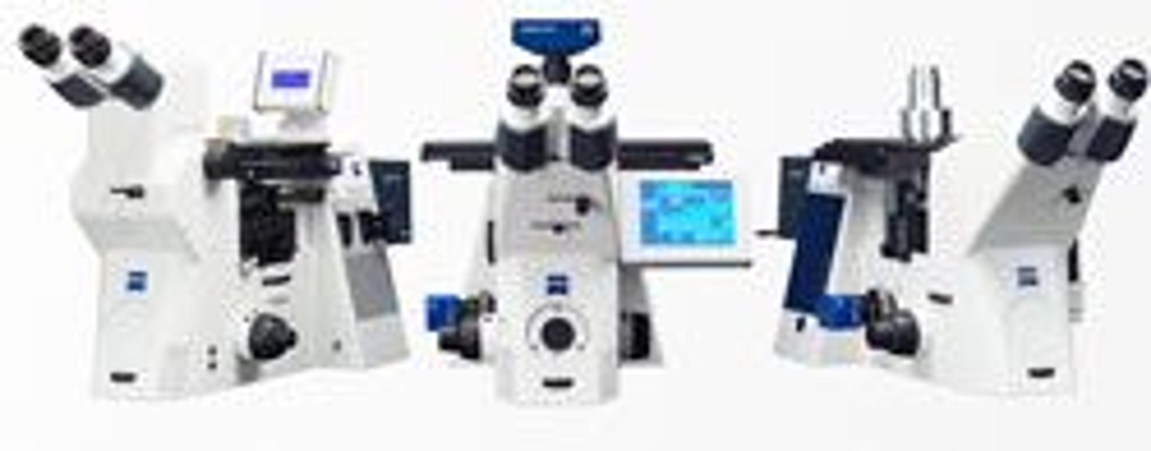
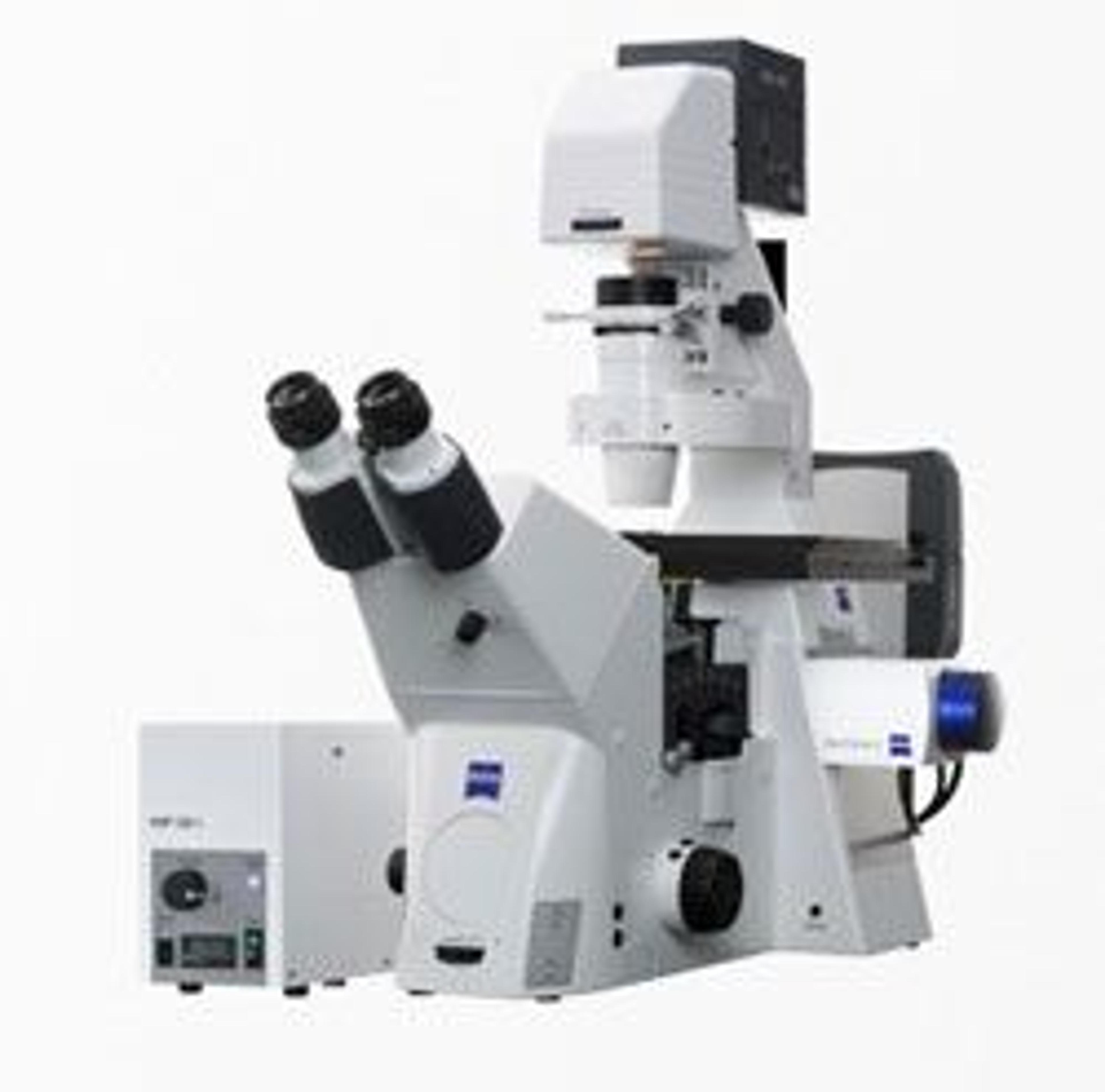
The Axio Observer family of light microscopes provides microscopy solutions for both biological and material science. The Axio Observer Live Cell microscope uses state-of-the-art LED illumination along with a variety of imaging modalities to suit the needs of the researcher. Additionally, increased automation, focusing, and image autocorrection features will allow for efficient sample analysis. The Axio Imager 2 microscope for material research allows for dependable, reproducible results for many quality control and process control needs. The four different stand versions allow for customization with options such as particle analysis, correlative microscopy, or upgradeable lenses. Also, the many contrast choices allow for the analysis of many different materials and surfaces.
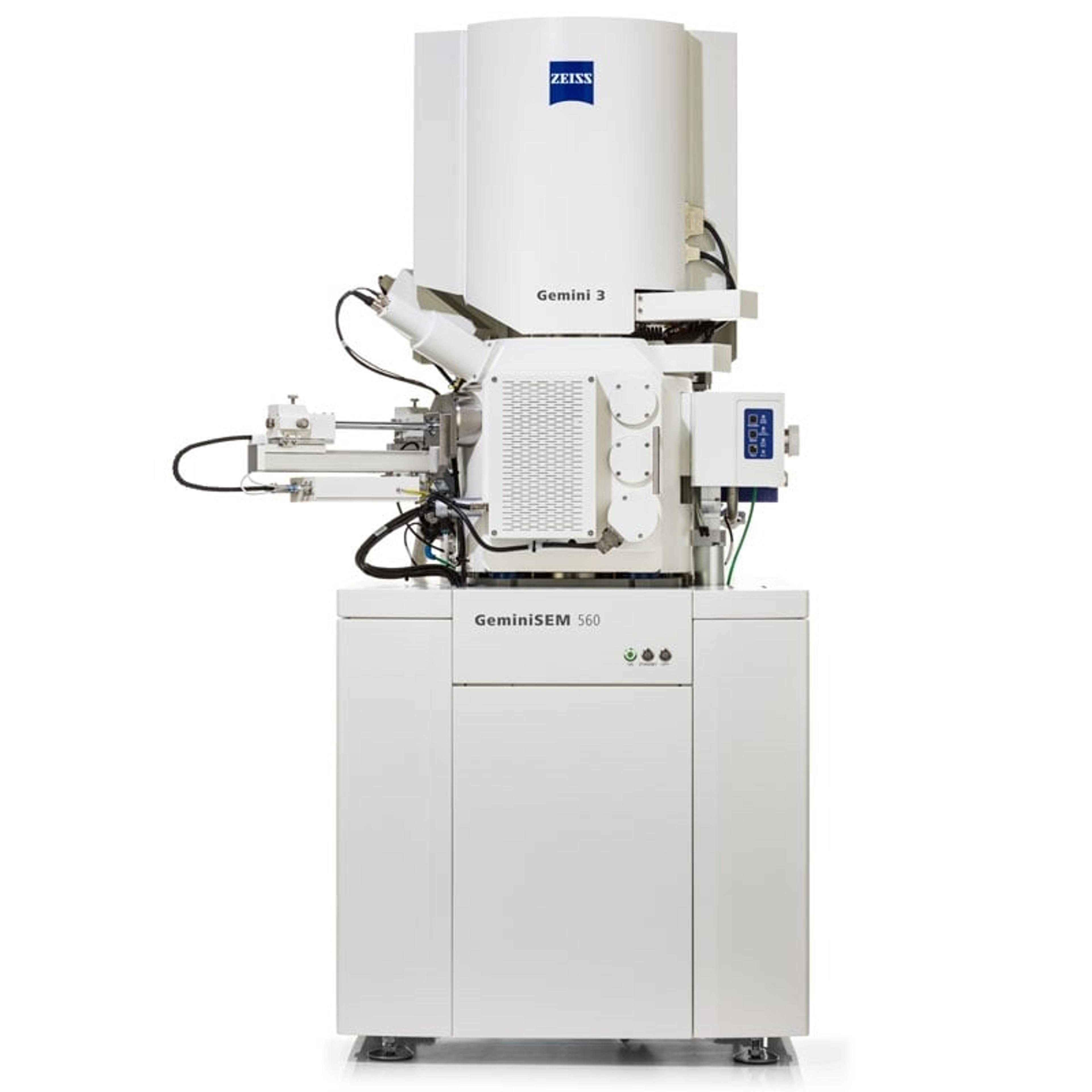
Booth visitors will also have an opportunity to view the GeminiSEM scanning electron microscope with its novel optical design. This microscope features a Nano-twin lens that produces high-quality images at lower voltages. With sub-nanometer resolution, even in variable pressure mode, crisp, high-contrast images can be acquired for any sample using the GeminiSEM. Due to the proven Gemini optical technology, even non-conducting samples can be used to produce high quality images. Scanning electron microscope enthusiasts will also be able to view the Video Station for the award-winning MultiSEM microscope, allowing for a better user interface and sample viewing experience.
Another product highlight will be the Primotech POL light microscope, a useful tool both for teaching and materials analysis. Using the innovative Matscope app, data from the Primotech microscope can be shared and accessed by multiple users, connecting many people along with numerous microscopes. All connected microscopes can be accessed via the app, allowing for a seamless data collection and analysis experiences. Moreover, the Primotech POL allows for easy analysis with reproducible results in the field of material science and analysis.
The Stemi 508 stereo microscope will also be on display at the ZEISS booth. This microscope features a generous 8:1 zoom and is meant to handle heavy workloads. In addition to the large zoom, the Stemi 508 has a 36 mm object field that allows crisp magnification of details up to 50x. This microscope is ergonomically designed to provide a comfortable working experience for everyday tasks with a low viewing angle of 35°. With four customizable stand configurations, the Stemi 508 can easily be outfitted for many applications.
With a fully digital platform and interface, the Smartzoom 5 microscope is ideal for many industrial quality assurance and quality control applications. Its simple operation and integrated QA/QC tools ensures that quality results are produced with every use, even among untrained users. The digital tools ensure smart workflow and smart output, providing a reliable solution for varied industrial needs.