ZEISS expands its portfolio of microscope cameras
Four new ZEISS Axiocam models are introduced for light microscopy applications
22 Nov 2019Product news
ZEISS has introduced four new high-quality CMOS cameras for digital imaging in light microscopy. These cameras complete the portfolio of proven ZEISS Axiocam models which stand for excellent performance in demanding microscopy applications.
The new microscope cameras ZEISS Axiocam 705 color and 712 color deliver the best possible image quality for histology, pathology or material research and analysis, thanks to excellent color rendition and greatly improved dynamic range. ZEISS Axiocam 705 mono and 712 mono are ideal for demanding fluorescence live-cell imaging with fast frame rates and high dynamic range. Also, their extended near-IR sensitivity allows for deeper insights into sample structures.
New high-quality sensors are boosting the cameras’ performance
Demanding microscopy applications call for a combination of excellent contrast, resolution, dynamic range, sensitivity and readout speed. In all these aspects, the performance of the four new ZEISS Axiocam microscope cameras benefits greatly from their new high-quality CMOS sensors. Small 3.45 µm pixels and low noise levels in combination with the fast USB 3.0 platform enable researchers to carry out extremely fast imaging experiments while maintaining excellent signal quality. Also, the new cameras’ global shutter architecture allows capturing dynamic samples without creating motion artifacts.
ZEISS Axiocam 705 mono and ZEISS Axiocam 705 color are 5-megapixel cameras optimized for fast speed and high dynamic range. ZEISS Axiocam 712 mono and ZEISS Axiocam 712 color are ideal for the acquisition of large samples with 12 megapixels, thereby greatly reducing the need for stitching.
Do you use Zeiss products in your lab? Write a review today for your chance to win a $400 Amazon gift card>>
Related products
Request Quote for All Products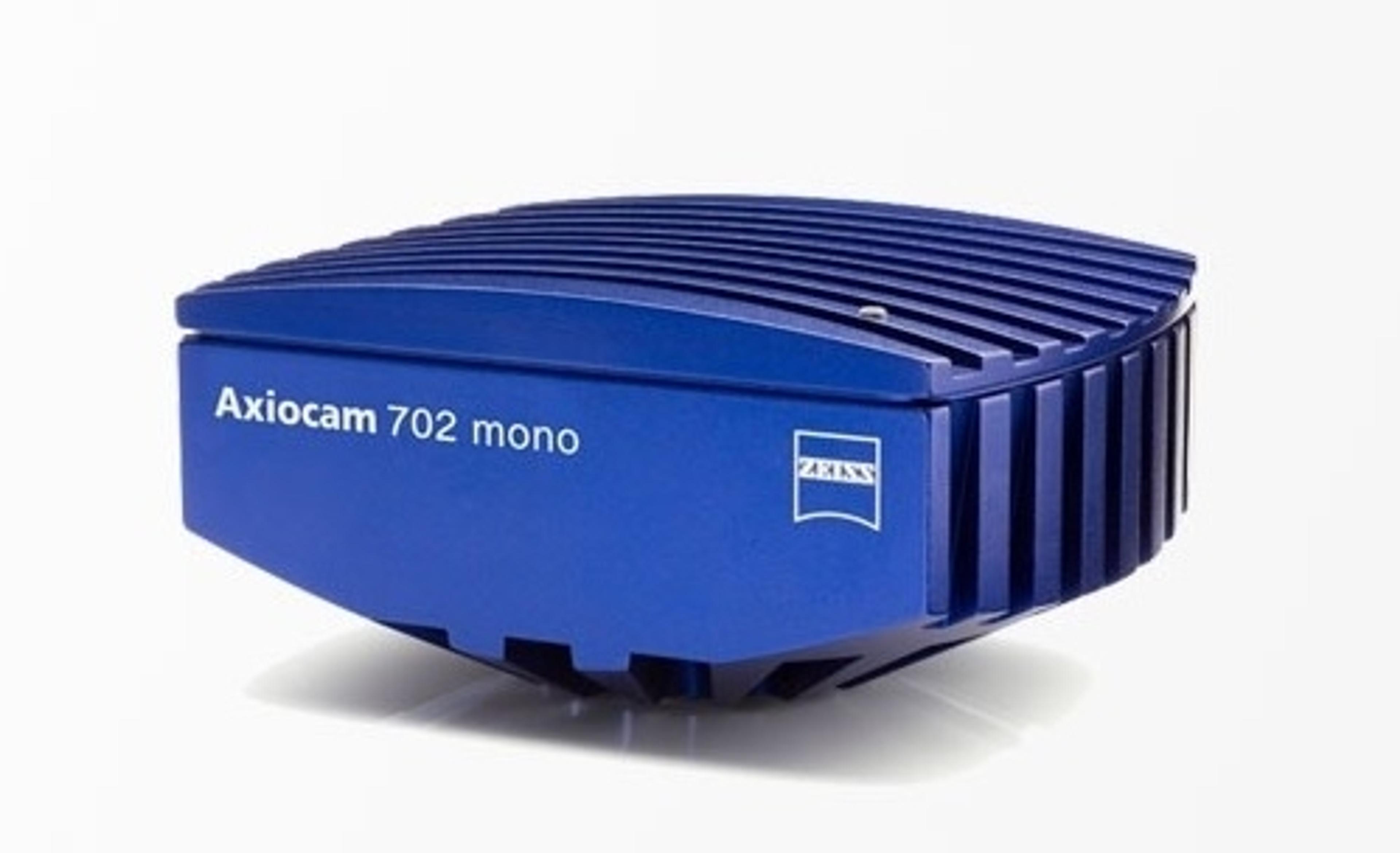
ZEISS Axiocam 702 mono
ZEISS Research Microscopy SolutionsYour 2.3 Megapixel Microscope Camera for Fast Low Light and Live Cell Imaging.

ZEISS Axiocam 512 color
ZEISS Research Microscopy SolutionsYour 12 Megapixel Microscope Camera for Imaging of Large Sample Areas.