Years on, the Nucleus Still Looks its Best in Red
21 Nov 2018
Product news
The far-red cell permeant DNA dye DRAQ5™ is now cited in more than 7600 peer-reviewed articles, heavily represented by top-ranked journals. Specifically, it has been widely used as a nuclear counterstain in high content screening (HCS) and associated drug discovery and development for more than a decade across Pharma’, CRO and academic labs and is backed by 200 independent customer reviews, as moderated by SelectScience.
To provide a new resource, the most recent of these papers have now been reviewed to include a brief snapshot of the topic, the platform used and details of the technique relevant to the use of DRAQ5™. Additionally, the resource signposts to useful book chapters. The resource can be found here: https://biostatusblog.blogspot.com/2018/11/DRAQ5DDrefs2017-2018.html
If you can’t find what you’re looking for there, BioStatus prides itself on providing front-to-back technical support, based on its many years of interactions with the drug discovery community and its own R&D programme, accessible directly from the website.
DRAQ5™ gives clear nuclear segmentation but also a useful secondary cytoplasmic signal for demarcation of the cytoplasmic envelope based on the real signal rather than reliance upon pixel dilation from the nuclear mask which aids signal quantification and observations of translocation events and morphometric indication of idiosyncratic toxicology. It can be used in both live- or fixed-cell endpoints and is spectrally separated from GFP and most vis-range chromophores. Because it is not excited by the UV wavelengths problems of compound interference are avoided. Remarkable chemical and photo- stability permit longer unattended operation on an automation deck in full-library screens. Importantly too, DRAQ5™ is cross-platform compatible for HCS imaging platforms, flow cytometers, imaging flow cytometers and fluorescence microscopes to simplify assay validation and transfer from bench-scale. A 1 ml pack of DRAQ5™ is sufficient for 100,000 wells (1536-mtp) or 33,000 wells (384-mtp) meaning its many benefits come at a small investment per datapoint.
Have you used DRAQ5 in your research? Write a review today for your chance to win a $400 Amazon Gift Card.
Related products
Request Quote for All Products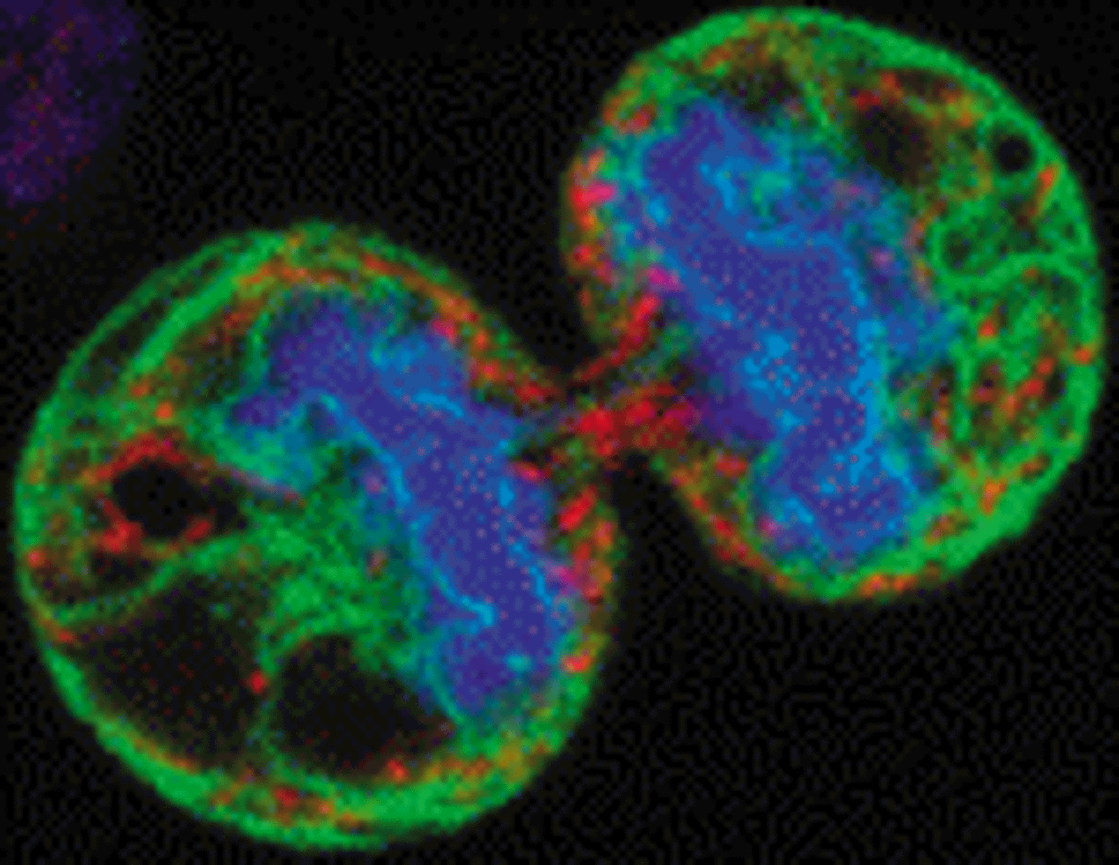
DRAQ5
Biostatus LimitedThe proven far-red probe for live or fixed cells, spectrally compatible with UV / Vis-range fluors.