Unraveling the tumor microenvironment of pancreatic cancer
Dr. Julienne Carstens highlights the critical role of spatial biology in understanding the metastatic progression of PDAC and advancing personalized immunotherapy
15 Jan 2024Editorial article

Dr. Julienne Carstens, Assistant Professor at the University of Alabama in Birmingham
Pancreatic ductal adenocarcinoma (PDAC) is the most common and deadly malignancy arising from the pancreas. It is therapeutically resistant, metastatic, and defined by a vast heterogeneous tumor microenvironment (TME) which is estimated to comprise up to 90% of the tumor volume in addition to the cancer cells themselves. Due to its extremely infiltrative nature and rapid tumor spread, only 5–10% of PDAC patients survive for five years or longer1. Investigating the cellular and molecular drivers of pancreatic cancer onset and metastasis is thus crucial to understanding the resistance of PDAC and developing therapeutic strategies to overcome it.
“Most patients with PDAC are diagnosed once the cancer has already disseminated throughout the body, meaning even if they undergo pancreatic resection to remove the cancer, they can relapse relatively quickly,” Dr. Julienne Carstens tells SelectScience®. “Understanding just what pancreatic cancer does when it's no longer in the pancreas is an open question.”
Dr. Carstens is an Assistant Professor at the University of Alabama in Birmingham, where she and her team are working to identify the driving forces behind PDAC progression, including both cancer intrinsic factors as well as the role of the tumor microenvironment. Using genetically engineered mouse models and patient samples, her team is combining spatial multiplex immunofluorescence technologies with multi-omics approaches to investigate the relationship between different cell populations in PDAC, with the ultimate goal to develop translational diagnostic biomarkers and enable targeted therapeutic intervention.
Who’s who in the TME
One of the key challenges presented by PDAC is understanding the composition of its unique tumor microenvironment. “We have these big buckets; fibroblasts, immune cells, pancreatic stellate cells, adipocytes, and extracellular matrix, but we still don’t know what all the cellular and noncellular components are,” shares Dr. Carstens. “A large majority of the research community are trying to understand what’s in the TME, the role and the function of all these individual cells, and how their interactions change when their environment is altered.”
Dr. Carstens likens the intricate network of the TME to human relationships on a microscopic scale. “Everything could be going really well in a neighborhood until you put in one bad actor, and everybody starts changing,” she says. “Some people may be passive, while others become highly aggressive and confront the bad actor. It’s about identifying who these people are and how they behave under different contexts.”
“However, unlike studying people, you can’t put a camera in the neighborhood and just watch what's going on,” she adds. Instead, her team is utilizing spatial multiplex immunofluorescence, which through the detection of multiple proteins by specific antibodies can provide a detailed picture of cell diversity, co-expression patterns, cellular interactions, and tissue architecture from a single tissue section. “Multiplex immunofluorescence allows us within a single snapshot to identify dozens of distinct cell populations and their activities in different tissues and under different conditions,” explains Dr. Carstens. “We’re now starting to get a script of not only the actors involved in PDAC but their interactions with each other and the role they play.”
Highly multiplexed immunofluorescence with streamlined optimization
For this work, Dr. Carstens and her team are using the Lunaphore COMET™, a fully automated hyperplex immunofluorescence platform capable of detecting 40 different spatial markers in each automated run on a tissue slide. According to Dr. Carstens, one of the key benefits of the platform is its scalability, particularly in terms of throughput, reliability, and reproducibility. “This allows us to start asking questions across multiple experiments and layer insights with different omics data, such as transcriptomics, genomics, lipidomics, and glycomics,” she says, adding that while many platforms are restricted to one sample, the COMET™ enables four slides to be processed simultaneously, which is especially advantageous when dealing with a full cohort of patient, archival, or experimental mouse samples.

The COMET™ can be used with any commercially available label-free antibody, while simultaneous optimization of multiple markers enables researchers to quickly establish reproducible assays without wasting precious samples and reagents.
While Dr. Carstens and her team are running the COMET™within a core facility using dedicated technicians, she notes that this is not essential given the instrument’s built-in protocols and intuitive operation. “One of the things that I really enjoy about Lunaphore is that they care very much about optimization,” she shares. “The instrument has pre-defined optimization criteria and guides users on how to optimize markers and imaging conditions as well as which types of optimizations they need to perform. This really lowers the bar for any user to get robust, reliable work.”
The COMET™ employs a sequential immunofluorescence (seqIF™) approach using patented microfluidic technology to precisely control cycles of staining, imaging, and elution. A gentle elution procedure is designed to optimize the preservation of tissue morphology and epitope stability between cycles, enabling multiple runs to be performed on the same slides – which is particularly important when dealing with precious samples. “With metastatic cancer, sometimes all you get is a core biopsy, if that, but using this technique many questions can be addressed on one slide,” says Dr. Carstens. “This is vital to be able to honor the contribution of patients and get as much understanding out of their samples as we possibly can.”
Clinical hurdles
Although researchers such as Dr. Carstens are already pushing the boundaries of discovery with spatial multiplex immunofluorescence, the road toward its clinical implementation is still in its infancy. “The bar for using pathology imaging as a diagnostic tool has already been met for immunohistochemistry (IHC), but the criteria for multiplex immunofluorescence will be even more stringent,” shares Dr. Carstens. “It will be essential to demonstrate that there are clinically significant aspects that can only be assessed through this approach. For example, if a unique cell type within a specific radius around a cancer is found to correlate with the tumor's susceptibility to a specific therapy – leading to a cure for those patients – it provides strong evidence for adopting this method as a diagnostic tool.”
“As a biologist, I envision continuing to use the technique to explore various relationships to discover what is truly important,” she adds. “However, hyperplexing may not be clinically necessary, as we may eventually identify and narrow down essential relationships that can be assessed by other means.”
The path to personalized immunotherapy
Current therapeutics have so far yielded limited success against PDAC, and it is expected to become the second leading cause of global cancer-related mortality in the near future1. “A pressing issue is that there’s currently no reliable way to determine if a particular chemotherapy regimen will be effective for a particular patient,” shares Dr. Carstens. “Moreover, due to the aggressive nature of this cancer, patients often don’t have the option of an initial treatment failing.”
This challenge is prompting ongoing efforts to define biomarkers that could aid patient stratification and tailor treatment selection to individual patients. “There is huge potential in understanding the biology of pancreatic cancer in a way where we could stratify patients for current chemotherapies and give them the best treatment the first time round,” she says. “Understanding the heterogeneity and the function of the microenvironment will be essential to these efforts.”
Looking ahead, Dr. Carstens is optimistic about the future of this research. “I think with the current level of support and momentum, coupled with technological breakthroughs and continuous improvements in model systems, we’re going to start to really understand PDAC biology, appreciate its complexity, and then reduce it down into something we can translate to the clinic. That's what I'm most excited about, and what our lab is focused on achieving,” she concludes.
References
Rahib L, et al., Projecting cancer incidence and deaths to 2030: The unexpected burden of thyroid, liver, and pancreas cancers in the United States. Cancer Res (2014).
Related products
Request Quote for All Products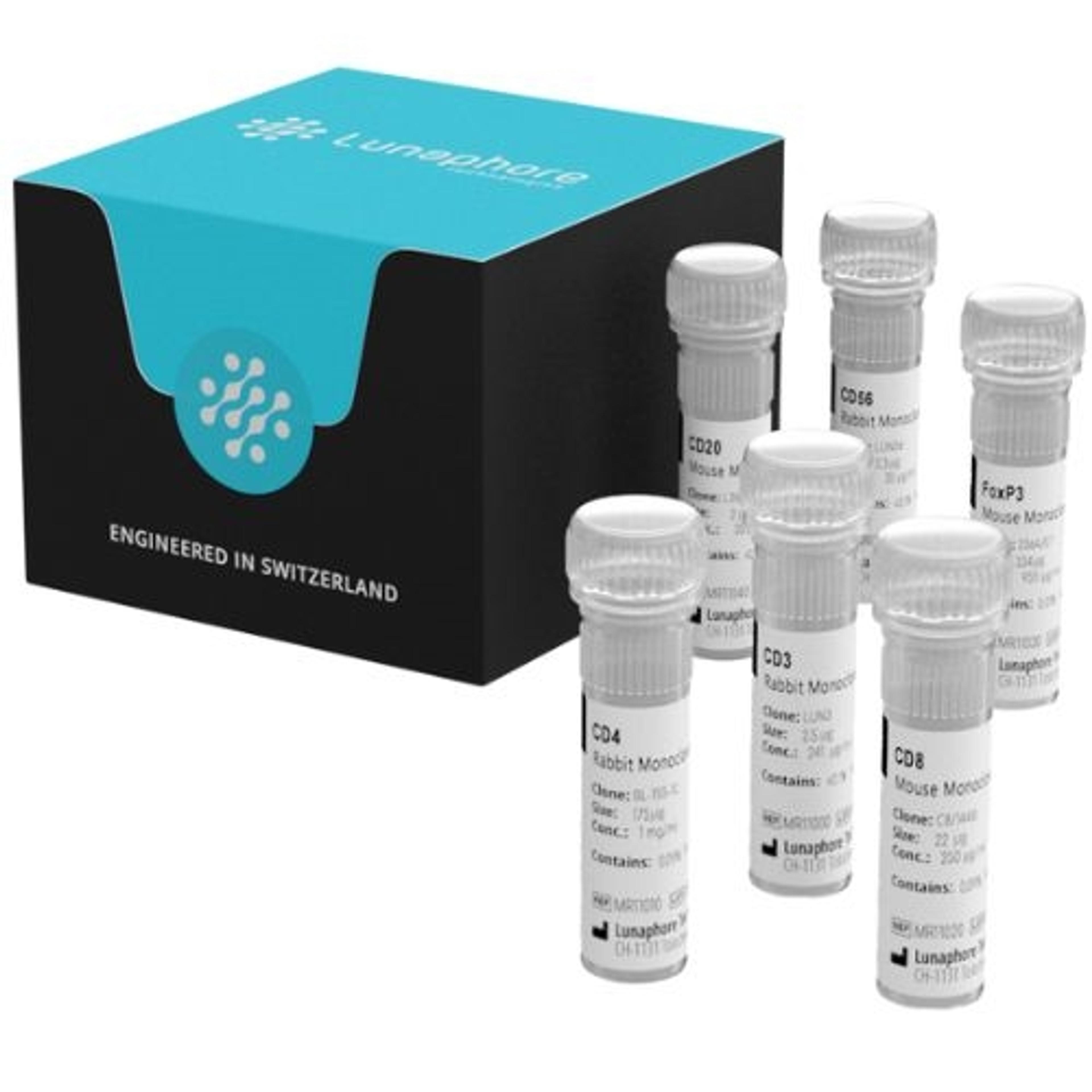
SPYRE Antibody Panels
Lunaphore TechnologiesTools to acelerate biomarker identification and validation
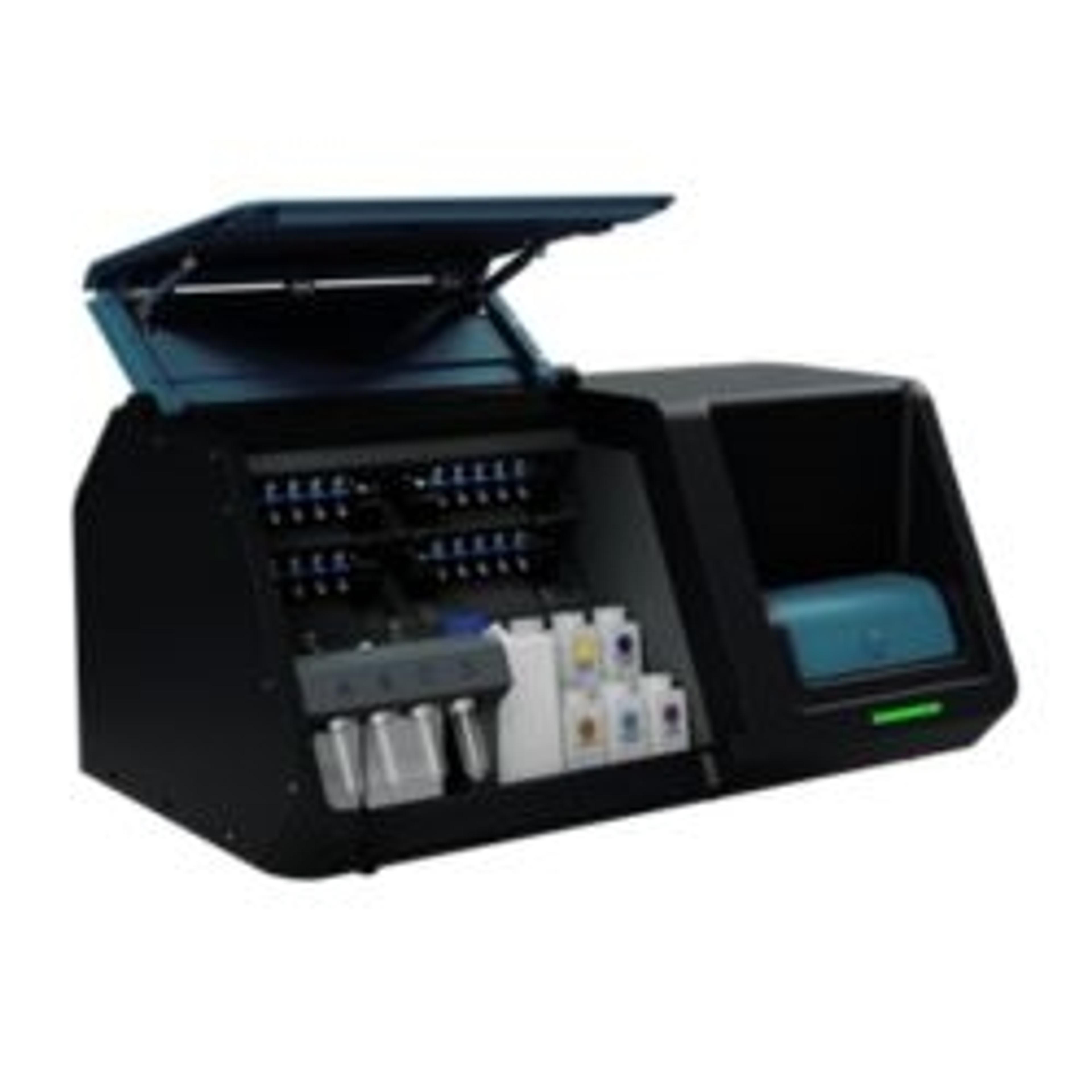
COMET
Lunaphore TechnologiesCOMET™ is a fully automated, high-throughput, hyperplex platform for the analysis of Formalin-Fixed, Paraffin-Embedded (FFPE), and frozen tissues.