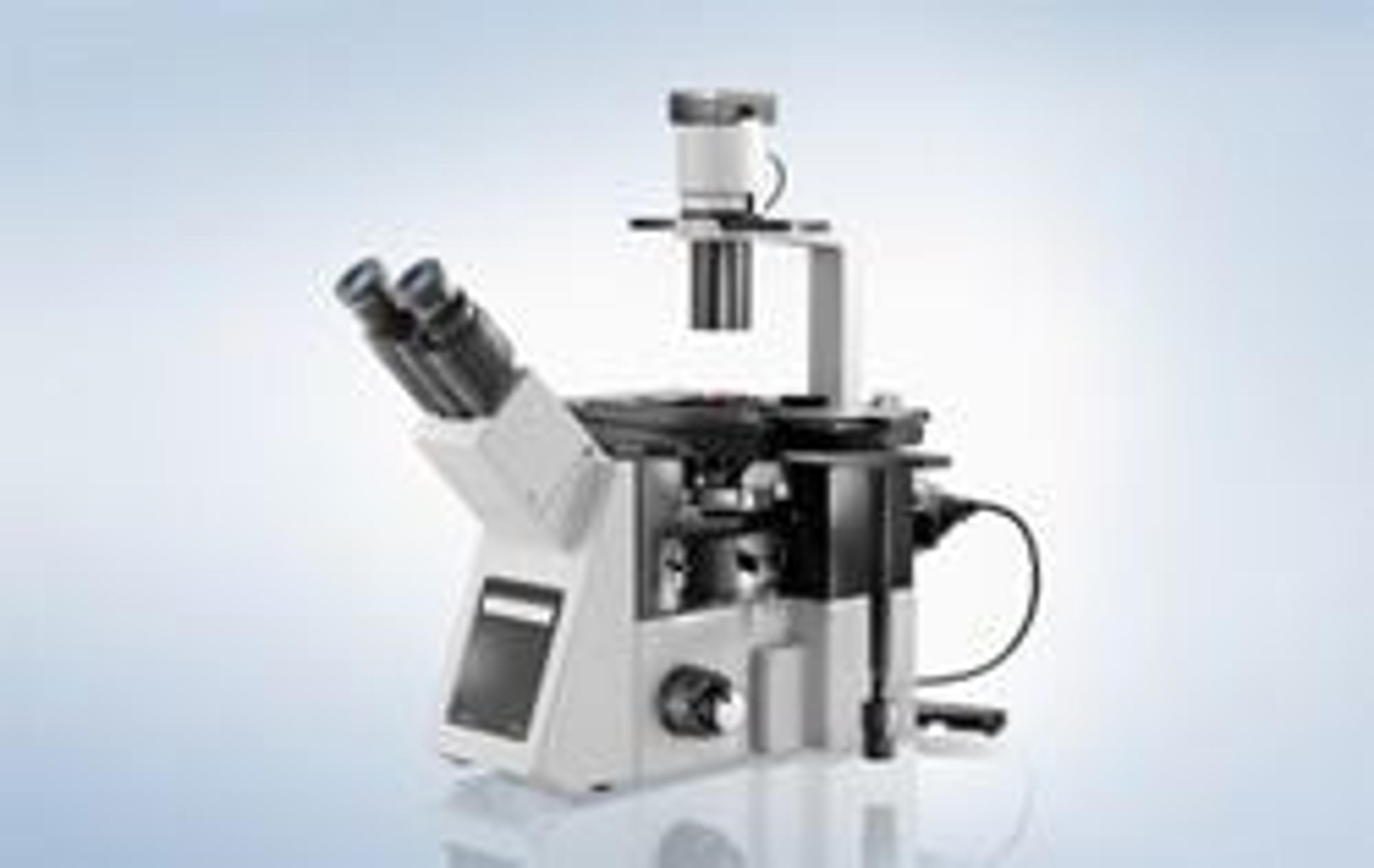
The New Olympus IX53 Inverted Microscope System For Frequent and Comfortable Routine Use
19 Sept 2012Product news
Olympus has released the new IX53 inverted microscope system for comfortable and constant use in the laboratory. Designed from the ground up to be the finest microscope available for routine analysis, the new system combines ergonomic operation with high quality optics in a cost-effective package.
Capable of bright-field, phase-contrast and fluorescence imaging, the IX53 offers a wide range of documentation and even research options. In particular, a long working distance condenser makes rapid, frequent Petri dish observations quick and easy, ensuring that the IX53 is the ideal choice for frequent, regular use, from checking cell culture plates through to the rapid screening of clinical samples.
The new IX3 series of inverted microscope systems have been designed based on extensive customer feedback to provide unrivalled ease-of-use and application flexibility. In particular, the highly-stable and robust IX53 is ideal for continual use in research and clinical laboratory environments, as it is exceptionally easy-to-use and has an ergonomic, low stage. The stage also utilizes a unique plate positioning system for easy sample tracking, in which user-defined position limits can be applied to immobilize the stage and sample. This ensures that the observation position is not inadvertently lost during operations such as reagent application, and makes it possible to transfer 35 mm dishes from the stage to an incubator and back, while still maintaining the correct field of view.
The IX53 frame is built utilizing an innovative, novel deck design, so it can be quickly and easily upgraded to facilitate new application capabilities, such as multi-channel fluorescence imaging. The microscope system also integrates seamlessly with Olympus’s powerful and intuitive cellSens software, providing hassle-free imaging and documentation.
Focused on usability and reliability, the cost-effective IX53 delivers exceptional performance across a wide range of routine tasks, with the expandability required to accommodate the needs of tomorrow.