"The most versatile whole slide imaging system": MMI launches MMI CellScan
Researchers are now able to scan full resolution digital slides in any magnification
3 Dec 2019
Product news
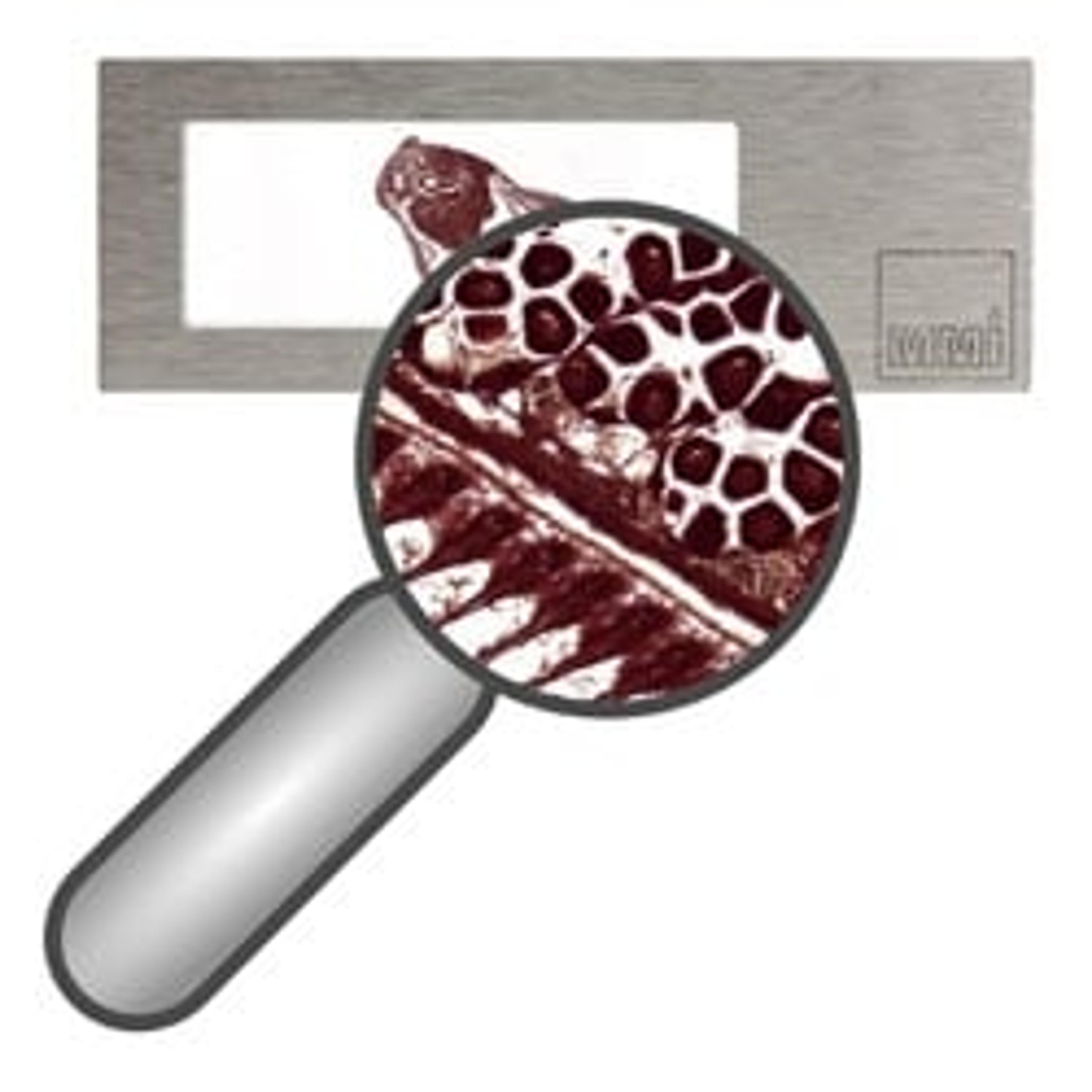
MMI officially launches their latest imaging solution at the Digital Pathology congress in London, UK. The MMI CellScan is a new stand-alone system for whole slide imaging. Researchers are now able to scan full resolution digital slides in any magnification and in any imaging modes, also in fluorescence.
For more than 25 years, MMI has been successfully developing and marketing unique instruments for selective single cell isolation, such as the MMI CellCut for laser microdissection. The MMI systems are based on inverted microscopes and thus support the flexibility provided by the microscope platforms.
Now, MMI launches the MMI CellScan, a stand-alone slide scanner based on the Nikon Ts2R microscope. The MMI CellScan can scan digital images using all objectives and imaging modes, even in fluorescence mode.
Scanned images, saved in the commonly used BigTIFF file format, can be quickly opened, instantaneously viewed and directly analyzed in their MMI CellViewer software. To facilitate remote work as well as interactive work in larger teams, they offer unlimited MMI CellViewer licenses. In addition, the BigTIFF files can also be imported into most other slide viewers.
The highly sensitive CMOS camera and the precise microscope stage enables for fast scanning rates. Moreover, the motorized z-drive promises to employ an individual focus map for highest image quality and to always focus on the right plane also in uneven samples.
With its small footprint, the MMI CellScan is designed to easily fit into your lab. In addition, the system can be also used as standard research microscope.
Dr. Stefan Niehren, CTO at MMI, is excited about the versatility of MMI’s newest solution: "The MMI CellScan is a very powerful and flexible tool exceeding the capabilities of standard slide scanners on the market today. Our CellScan is compatible with all objectives, with all standard imaging modules, with most Slide Viewers and, of course, with all MMI products. "
“In addition to our CellScanM module which can be uniquely combined with laser microdissection, we can now offer the standalone CellScan to provide Whole Slide Imaging for many more applications, for example in pathology but also in standard research”, highlights Prof. Dr. Stefan Seeger, CEO at MMI.
Do you want more of the latest science news straight to your inbox? Become a SelectScience member for free today>>