Stepping up The War on Cancer with New Technology

Learn how Dr Robert Hoffman is shifting the paradigm in cancer research.

17 Jan 2017
Emily Adam
Publishing / Media

Editorial article

 The iBox® Scientia™ Small Animal Imaging System offers three main advantages, Dr Hoffman's team found.

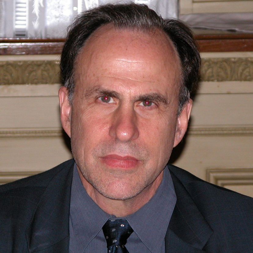
Dr Robert Hoffman, AntiCancer Inc.

Dr Robert Hoffman, founder of AntiCancer Inc., and Professor of Surgery at USCD, San Diego, told SelectScience® about recent advances in technology that have significantly impacted his cancer research. AntiCancer Inc. has employed iBox® Scientia™ Small Animal Imaging System produced by Analytik Jena US, formerly UVP LLC, to make several breakthroughs, at greater resolution, and at a fraction of the cost compared to the analytical tools commonly used in cancer research.

Dr Hoffman’s current research focus is on translational techniques - finding what works in the laboratory that can quickly be translated to benefit patients, whether that benefit comes from effective imaging systems, or developing strains of bacteria to directly target tumors. With a successful track record in cancer research dating back to the mid 1960s, Dr Hoffman’s efforts are now channeled into working with tumor-targeting bacteria, and other areas of cancer research.

Hoffman’s models represent a paradigm shift in cancer research. “We’re still treating solid tumors like a leukemia or a lymphoma. This is wrong as most solid tumor cells are at a non-dividing stage in their cell cycle and must be treated differently,” added Hoffman, “Cancer patients don’t respond to drugs as the cells in their tumors are mostly non-dividing, and therefore resistant to drugs.” Hoffman linked this ‘black box’ approach to hair loss, a common side effect of traditional cancer therapies. “Hair cells die because they’re always dividing. The bacteria employed in AntiCancer Inc.’s research attacks tumors and decoy the cancer cells to transition to the most sensitive part of the cell cycle."

AntiCancer Inc. uses high-resolution imaging to study single cells in the mouse model, together with micro surgery to implant the tumors. “Very few labs can do this. Imaging and microsurgery are very important to us.” “The iBox® Scientia™ Small Animal Imaging System offers us three main advantages,” said Dr Hoffman. “It gives us high resolution images, with high contrast, and at high magnification. It can even give us spectral separation – everything you need.” Dr Hoffman’s team has been able to study the activity of individual cancer cells non-invasively, using fluorescent proteins and the iBox®. Hoffman continued, “We have over two hundred different models of cancer. We can image inside a mouse using a fluorescent protein – and we can see through the skin – it’s not invasive.” Hoffman concluded, “We also found the iBox to be so cheap – you can buy an iBox® system for around a quarter of what we’d expect to pay for a single analyzer of an established brand. UVP has overcome what is a barrier to smaller labs – a better machine and at a quarter of the price, and with many different options.”

Alluding to his comments on the current ‘black box’ approach, Hoffman is frustrated for the future of cancer research. “I worry that the leaders are working in the wrong paradigm,” he added. “It will be a long and a big fight – it’ll take decades if not another century.” But Hoffman sees great opportunities in other areas. “I am excited about stem cells, we want to be able to regenerate everything.” Hoffman added, “With stem cells that come from the hair - everybody’s hair works. With these cells, we want to be able to regenerate the spinal cord, nerves, heart. And hair cells are accessible from every individual without having to worry about any immune problems, and they don’t make tumors.”

Read the latest stem research from Dr Hoffman’s group to learn more about how they’ve developed tumor-targeting bacteria to attack cancer in mouse models, image single cells in live mice, how hair-associated-pluripotent (HAP) stem cells repair nerves and differentiate into heart cells, how human tumor behaviour is replicated in a mouse and many other new technologies.

Do you use the iBox® Scientia™ Small Animal Imaging System? Tell us what you think, leave a review today.

Links

Tags

Cell Lines Stem Cells and Primary CellsPrimary cell cultures, established cell lines and stem cells are vital for <i>in vitro</i> and <i>ex vivo</i> experimentation. High-quality cells, optimized for your applications, alongside optimized cell substrates, growth medium and supplements, are critical for experimental success. Explore a range of cells suitable for your applications, including isogenic cell lines, competent cells, induced pluripotent stem cell (iPSC)-derived cell lines, fungal/bacterial/mammalian cell lines, stem cells and cancer cell lines. Find the best cells for your research in our peer-reviewed product directory: compare products, check customer reviews and receive pricing direct from manufacturers.Cellular PathologyCellular Pathology deals with the microscopic analysis of tissue samples and cells. Sample preparation and processing includes fixation, staining, sectioning and slide mounting, using equipment such microtomes and cryostats. In choosing immunohistochemistry and immunocytochemistry kits, consider chromogens, staining method, antibodies, microscopes and imaging.Protein PurificationProtein purification is a vital step in drug discovery, therapeutics, biotech and life science research. The purification process typically involves subcellular or membrane protein extraction with cell lysis kits, separation of proteins from cell debris by filtration or spin columns, and the isolation of proteins of interest from other proteins and impurities with affinity purification (including fusion protein tags and antibody binding proteins A, G and L), immunoprecipitation or chromatographic methods, such as ion exchange, size exclusion and immobilized metal affinity chromatography. All purification methods come in multiple formats for your laboratory needs, including agarose or magnetic beads, resins, columns and filter plates. Find the best protein purification equipment in our peer-reviewed product directory: compare products, check customer reviews and receive pricing direct from manufacturers.In Vivo Imaging Systems<i>In vivo</i> imaging systems, including pre-clinical imaging systems and medical imaging systems are used to non-invasively visualize and capture images of live animals and plants. Monitor the natural processes or diseases of your subjects using small-animal pre-clinical imaging systems, including single photon positron emission tomography (SPECT), positron emission tomography (PET), computed tomography (micro-CT), magnetic resonance imaging (MRI), X-ray radiography, ultrasound, fluorescence and bioluminescence imagers. Multimodal systems and software solutions are also available for correlative analysis of organ, tissue, cell, or molecular-level processes. Find the best in vivo imaging products in our peer-reviewed product directory: compare products, check customer reviews and receive pricing direct from manufacturers.Translational ResearchLive Cell ImagingLive cell imaging is the study of living cells using microscopes and high-content imaging systems. This technique provides in-depth insight into fast and complex biological processes, by allowing dynamic imaging of living cells instead of acquiring an individual image at a single point in time.Translational MedicineTumorsTumor research focuses on understanding abnormal cell growth that leads to cancer. Identifying biomarkers, studying tumor microenvironments, and developing targeted therapies are critical for advancing cancer treatment. Early detection and personalized treatment options are key to improving outcomes for patients. Browse our peer-reviewed product directory to explore tools for tumor research, diagnostics, and cancer therapies; compare products, read customer reviews, and get pricing directly from manufacturers.Cancer ResearchAlthough cancer is often referred to as a single condition, it actually consists of more than 100 different diseases. Microscopy, mass spectrometry, high throughput sequencing and flow cytometry are some of the most common techniques employed in cancer research labs.
Stepping up The War on Cancer with New Technology