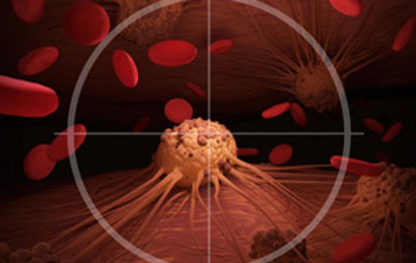
Spatial biology for cancer research: A powerful new tool
These resources share the latest progress and technological advances in spatial biology, which can be utilized to improve cancer research
17 Nov 2022
Editorial article
Improvements in spatial biology for cancer research techniques can support a range of different research areas. Developments can enhance imaging, spatial phenotyping, cell analysis, and general understanding of the biology of tumors and other tissues within their existing structures.
We’ve selected a range of resources that present the latest insights into the field of spatial biology for cancer research.
Bringing spatial biology to the clinic

In this article, spatial transcriptomics pioneer, Dr. Arutha Kulasinghe, shares his latest work utilizing spatial biology in cancer research to understand the underlying tumor biology, using an integrative multi-omics approach, and how it could inform therapy decisions.
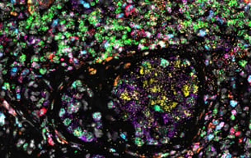
Predicting cancer immunotherapy response by highly multiplexed tumor imaging
The tumor microenvironment (TME) – the main site of tumor-immune cell interactions – crucially regulates antitumoral immunity and immunotherapy response. Watch this on-demand webinar to discover how highly multiplexed microscopy can be used to thoroughly characterize tumor microenvironment cell types, their spatial interactions, and the tumor architecture to predict patient response to immunotherapy in various cancers.
Spatial biology: Context matters

This video by Akoya Biosciences explains the importance and potential of spatial phenotyping in fields such as cancer, immunology and infectious disease research, and how imaging solutions can enable you to phenotype the full cellular diversity of a tissue sample, with spatial context, just like a single cell GPS.
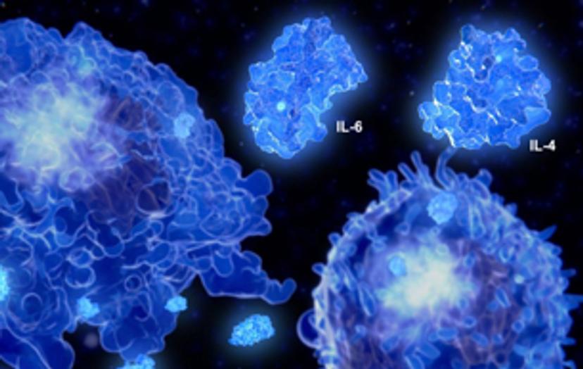
Taking a multi-omic multiplexing approach to spatial biology
In this free eBook, we present a series of case studies illustrating how a multi-omic multiplexing approach can be leveraged to gain a comprehensive view of spatial biology, including a multiplexed in situ transcriptomic method for the spatial mapping of target genes in highly complex and heterogenous FFPE tumor tissues, and much more.
Spatial phenotyping: Bringing the power of spatial biology to cancer research and therapy
In this whitepaper, Leinco Technologies explores how spatial-omics, especially spatial phenotyping, is revolutionizing translational medicine for the treatment of cancer, and the key role played by high-quality antibodies in the generation of data that is reliable and reproducible.
The power of spatial biology: A microscopy guide

Location is key to understanding biological mechanisms, from the inner workings of subcellular components to how cells form and interact across normal and diseased tissues. Many application areas are seeing a growing trend toward spatial biology, which uses transcriptomics, imaging, and other approaches to put dissociated cellular information into spatial context. In this free eBook, explore key microscopy techniques across the spatial biology workflow. Plus, we look 'under the microscope' at how these can be applied to study a wide range of spatial biology questions and gain expert insight into the imaging solutions designed to meet a variety of different research needs and priorities.
The Orion Platform for Spatial Biology

In this product brochure, discover the Orion™ novel technology and services platform offering a fast path to whole slide, high-plex imaging. Combining speed and resolution, the Orion platform enables comprehensive phenotypic profiling and characterization of tissue architecture, tumor heterogeneity, and the immune response for whole sections in hours.