sCMOS Camera with High Sensitivity, High Resolution and Fast Readout
16 Jan 2012Product news
Hamamatsu Photonics has introduced the new ORCA-Flash4.0 sCMOS camera. In recent years, trying to detect low level or fast fluorescence signals has proved challenging and an EM-CCD camera was commonly used, however, with the arrival of the ORCA-Flash4.0 that situation has now changed. The ORCA-Flash4.0 is the first camera to be able to deliver better signal-to-noise than EM-CCDs, cooled CCDs and first-generation sCMOS cameras for low-level, fast, fluorescence imaging.
The ORCA-Flash4.0’s high sensitivity means extreme versatility. This camera covers a wide range of imaging needs, including super resolution microscopy, TIRF microscopy, live cell GFP, high-speed calcium ion imaging, FRET, real-time confocal microscopy and many more.
The ORCA-Flash4.0 has quantum efficiency values of over 70% at 600 nm and 50% at 750 nm and has only 1.3 electrons of read noise (at 100 full-resolution frames per second) with continuous high speed acquisition at full resolution. This unique combination of high quantum efficiency and low noise, in the absence of EM-CCD multiplicative noise, means that researchers are already pushing back the boundaries of what has been possible to image until now. With a 4.0 megapixel sCMOS sensor (6.5 μm pixel size), the ORCA-Flash4.0 is ideally suited for demanding microscopy applications.
Related products
Request Quote for All Products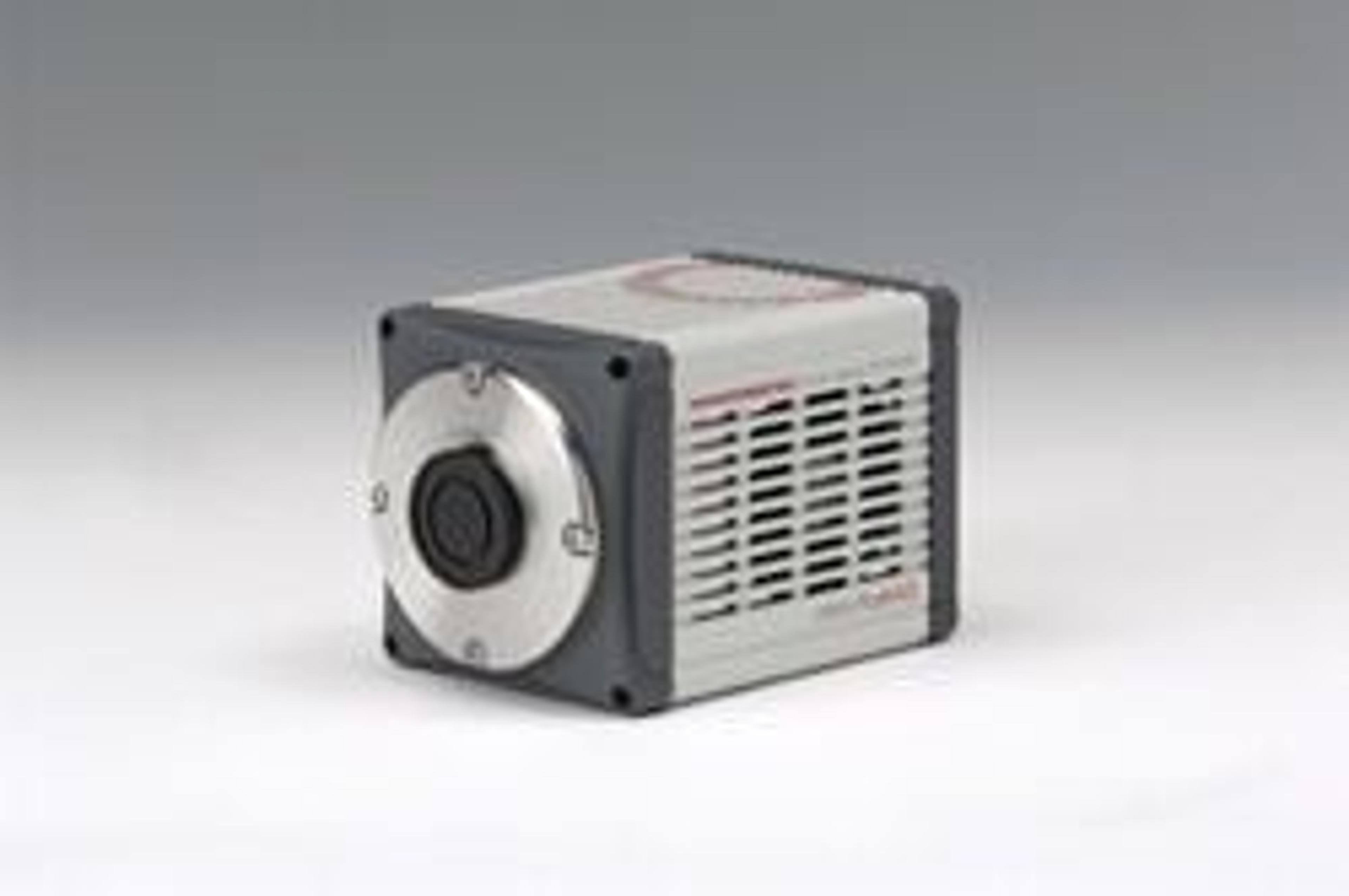
ORCA-Flash4.0 Camera
Hamamatsu Corporation Camera Products GroupHamamatsu’s brilliantly designed ORCA-Flash4.0 is truly a game changer in the world of scientific imaging. Built on a revolutionary new Gen II sCMOS detector, the ORCA-Flash4.0 is the first sCMOS camera that challenges the performance of all CCD, EM-CCD, and Gen I sCMOS cameras. With its combination of low noise and high quantum efficiency, the ORCA-Flash4.0 delivers unprecedented sensitivity as well as high dynamic range, blazing fast speeds, large field of view, and excellent resolution—all at once. Scientists have used EM-CCDs for very low-light, often high-speed imaging such as TIRF or spinning disk confocal, while they have relied on cooled CCDs for other fluorescence applications such as GFP or multichannel imaging. But the ORCA-Flash4.0 is changing all of that. Now one camera covers a wide range of imaging needs—including localization microscopy, TIRF microscopy, live cell GFP, high-speed calcium ion imaging, FRET, real-time confocal microscopy, and many more. The ORCA-Flash4.0 is a solid piece of camera engineering. It has optimized cooling and outstanding image uniformity across the entire sensor compared to cameras based on Gen I sCMOS technology. Other features include 16-bit A/D converter and multiple external trigger and timing output functions.Applications • Localization microscopy • TIRF microscopy • Live cell GFP • Time lapse fluorescence • Ratio imaging • FRET • High-speed calcium ion imaging • Real-time confocal microscopy • Light sheet microscopy • Morphology • Fluorescence in situ hybridization (FISH) For more information on the ORCA-Flash4.0 please visit our website or request more information at the top of the page.