Renishaw introduces breakthrough time-resolved Raman spectroscopy integration
TRRS leverages the faster interaction time of Raman scattering to distinguish the Raman signal from the fluorescence background
16 Jan 2026Product news
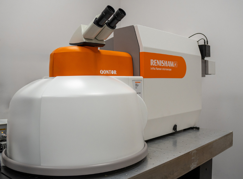
inVia™ confocal Raman microscope
Renishaw has launched its latest innovation in Raman spectroscopy: the integration of time-resolved Raman spectroscopy (TRRS) into its inVia™ confocal Raman microscope. This cutting-edge technique enables researchers and engineers to overcome the long-standing challenge of sample fluorescence, unlocking high-quality Raman spectra from samples previously deemed unmeasurable.
TRRS leverages the rapid interaction time of Raman scattered photons — occurring within picoseconds of laser excitation — to distinguish them from the slower fluorescence background. This technique employs a variety of state-of-the-art technological innovations including a newly developed single-photon avalanche diode (SPAD) array detector, designed and manufactured by Singular Photonics.
This sensor utilizes advanced CMOS SPAD technology and can detect individual photons and record their arrival time with 50 picosecond temporal precision. Historically, isolating the Raman photons from the fluorescence background has been complex, but with Renishaw’s proprietary algorithms, extracting Raman data is automatic, ensuring this advanced technique is accessible to a wide range of users.
The TRRS integration enables Raman analysis of a variety of challenging samples, such as food, oils and lubricants, and polymers (including those containing pigments and dyes).
Renishaw and Singular Photonics have been developing this technology, with multiple associated patents, for several years. This latest integration marks a significant milestone in Raman spectroscopy, making it possible to unlock chemical and structural information from real-world samples with high background fluorescence that were previously inaccessible.
Want the latest science news straight to your inbox? Become a SelectScience member for free today>>
Related products
Request Quote for All Products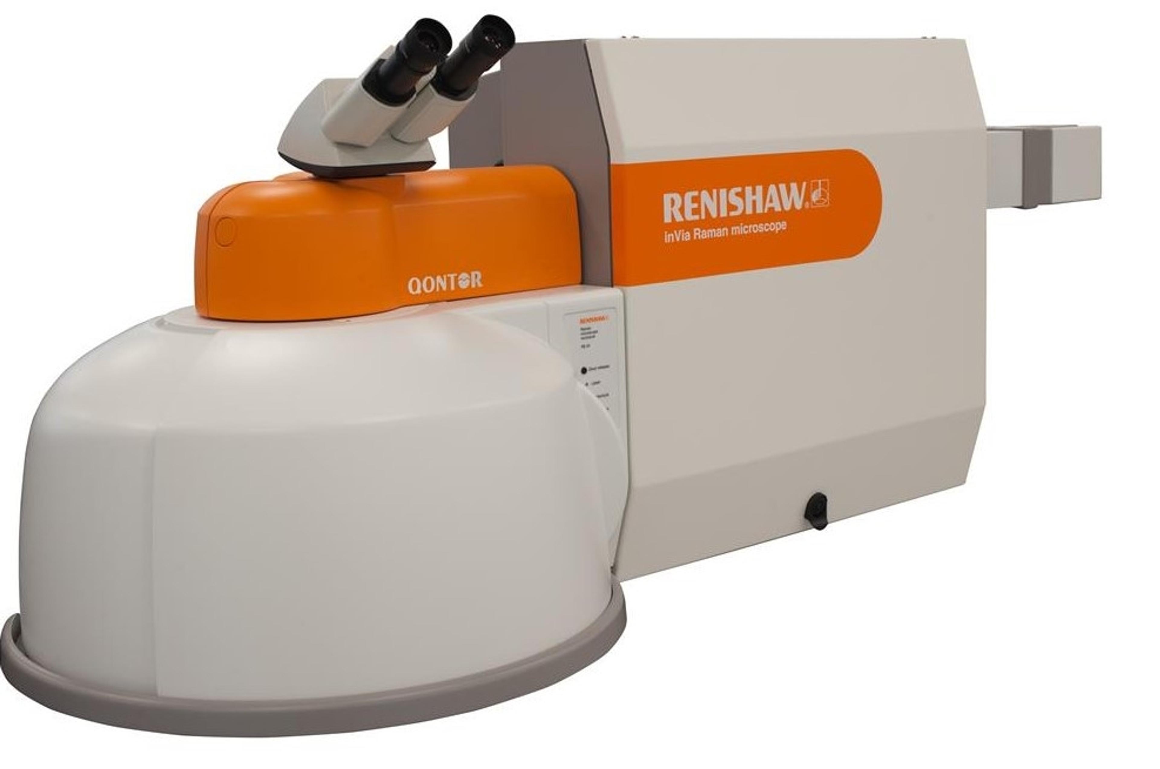
inVia™ Qontor® confocal Raman microscope
Renishaw plc.Designed with the latest LiveTrack™ focus tracking technology, the inVia Qontor enables users to analyse samples with uneven surfaces
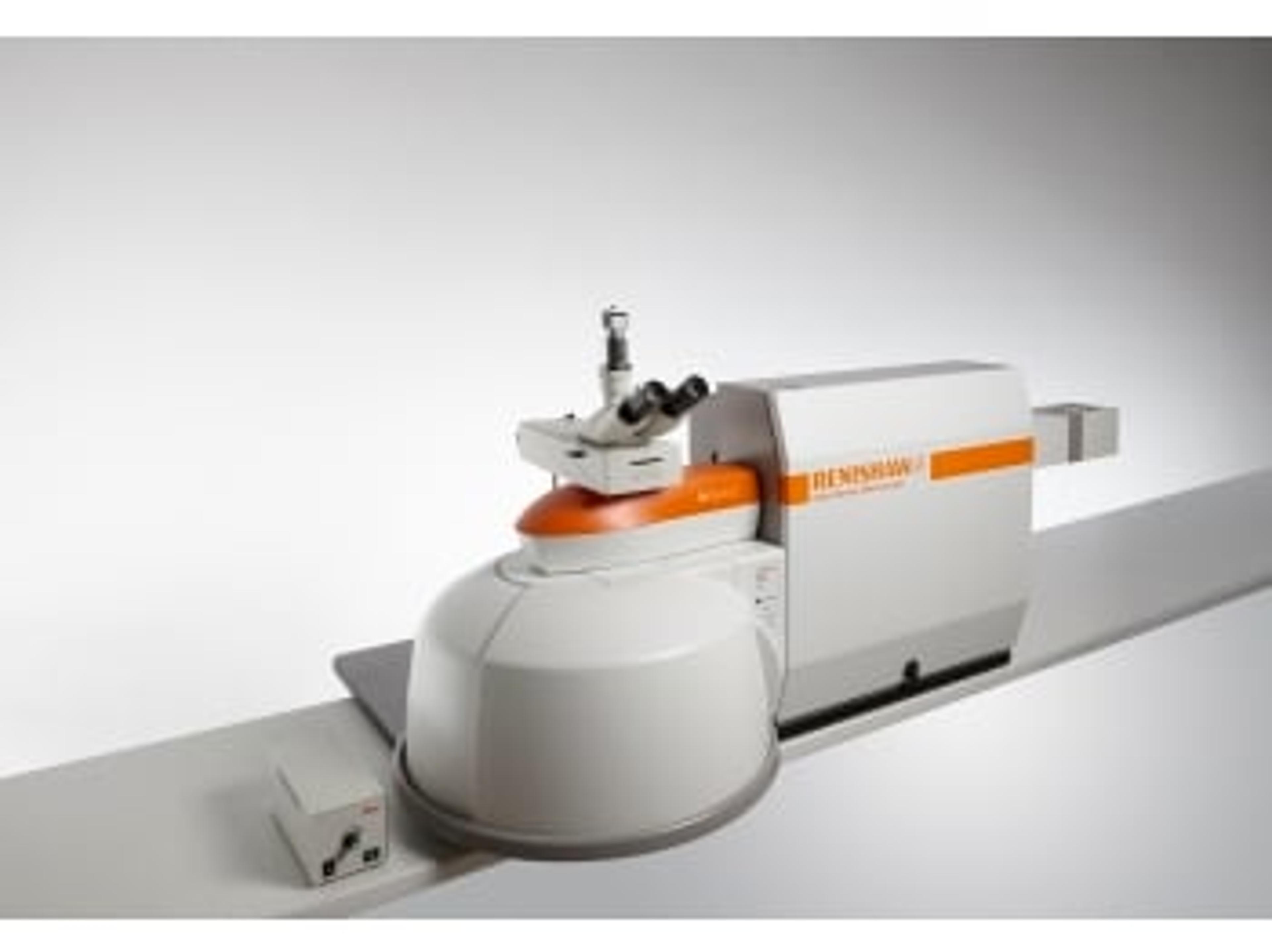
inVia InSpect confocal Raman microscope
Renishaw plc.Confocal Raman microscope optimised for use in forensic laboratories for trace analysis
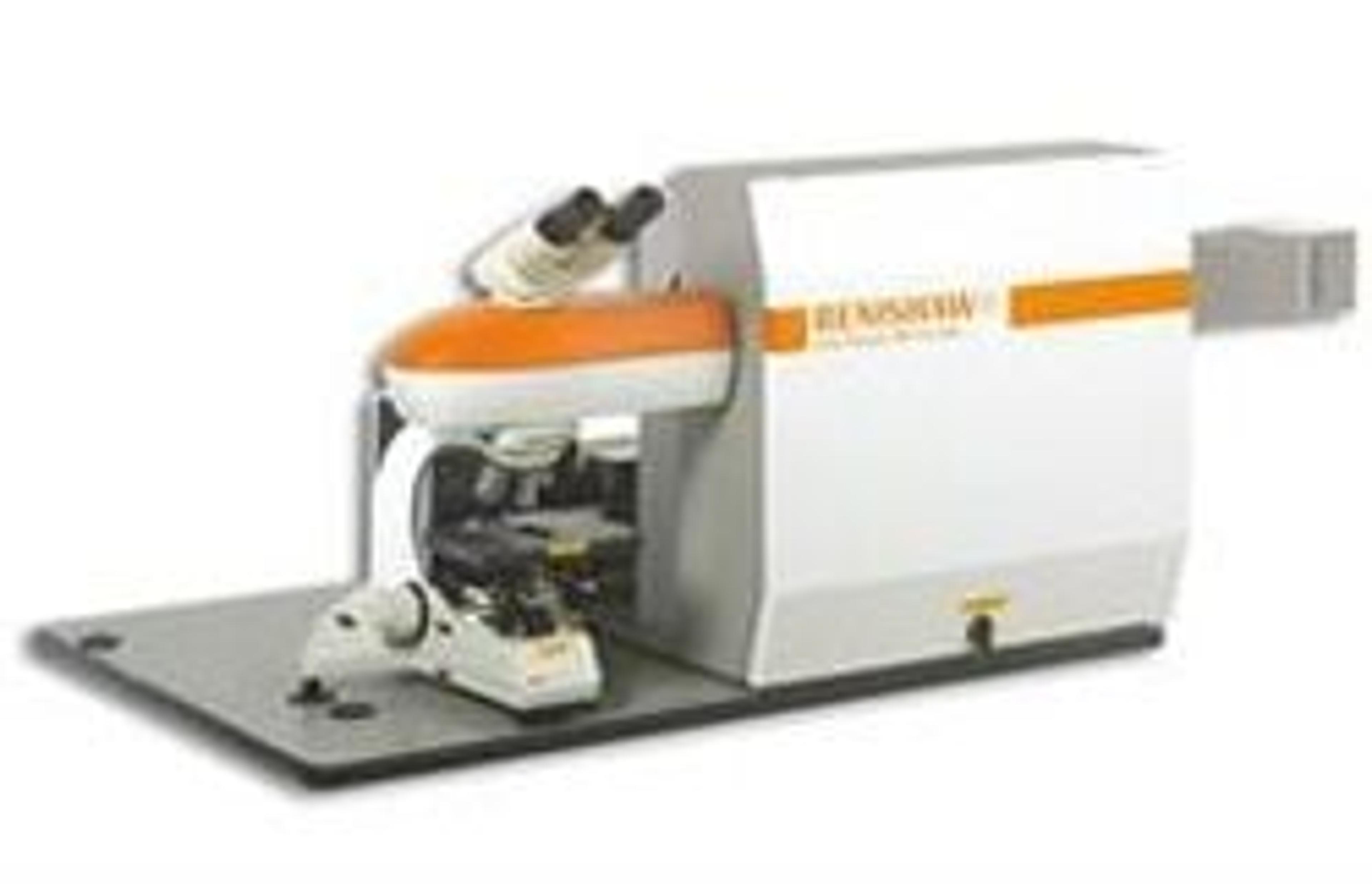
inVia Raman microscope
Renishaw plc.Since being launched, the Renishaw inVia Raman microscope has become the world's best selling research Raman system. The inVia Raman microscope combines simplicity of operation with the high performance and unparalleled flexibility for which Renishaw Raman systems are renowned. inVia Raman microscopes are high-sensitivity systems with integrated research grade microscopes, enabling high resolution confocal measurements. inVia Raman microscopes support multiple lasers, with automatic software switching of excitation wavelength.