Olympus Present Open Source Microscope Concept at FOM 2013
13 Mar 2013
Product news
Olympus presents various systems based on the unique, flexible IX3 inverted microscope frame series. The systems will highlight easy-to-use adaptability of the new microscope concept from routine observation through to advanced imaging techniques.
Olympus will exhibit their IX3 inverted microscope systems for live cell applications in booth 46-48 at the Focus on Microscopy 2013 (FOM) annual conference in Maastricht, Netherlands, 24th-27th of March. The new open source frame concept has been designed around an infinite light path made easily accessible with interchangeable optical modules that can be swapped in and out of the light path, enabling the creation of various distinct systems on one inverted microscope frame.
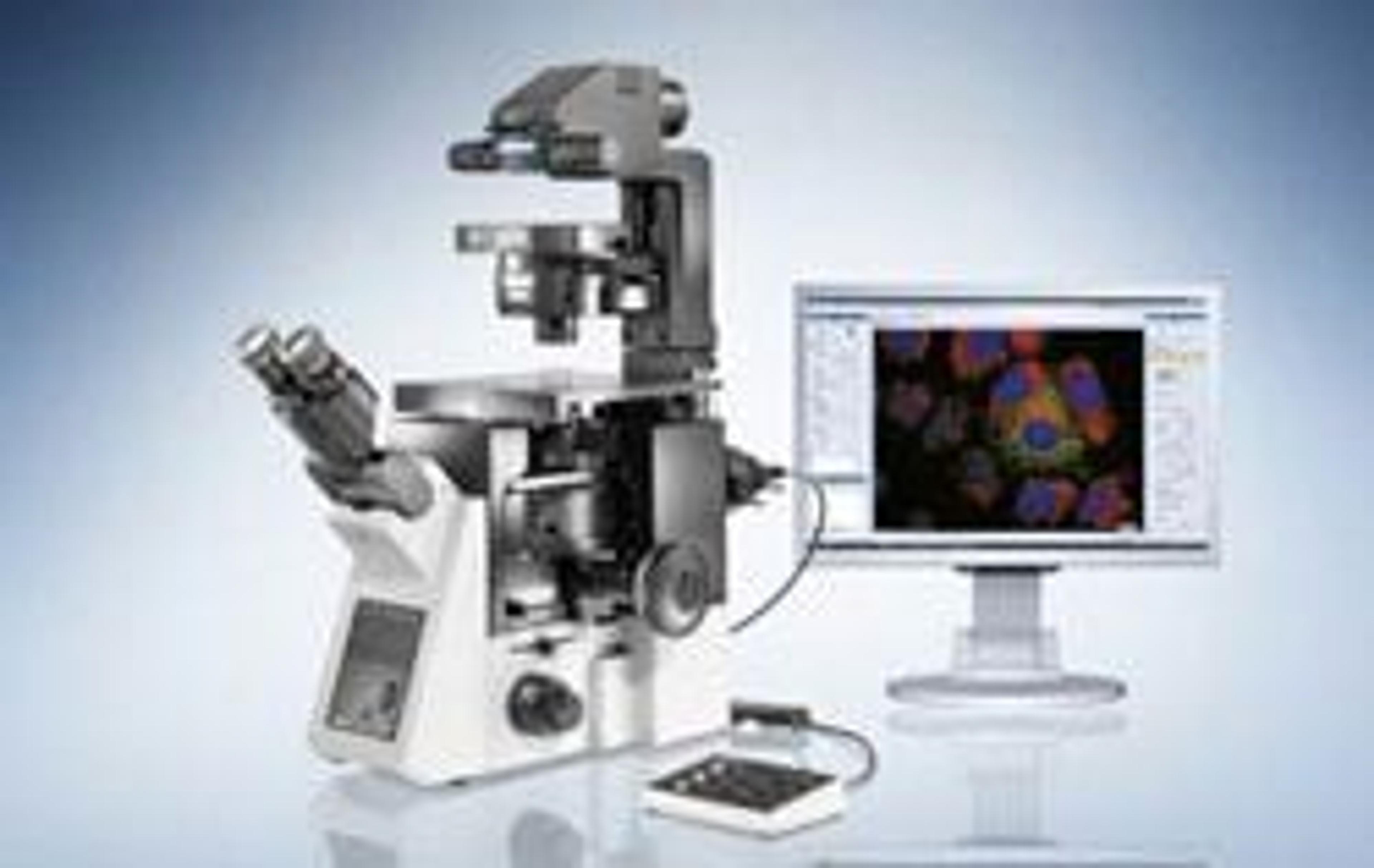
Presentations of example systems will include the IX73 with cellSens and LED illumination control for fast imaging, the xcellence imaging system with IX83 for total internal reflection fluorescence microscopy (TIRF), and the FluoView FV1200 with IX83 for confocal laser scanning microscopy.
FOM brings together global leaders within the microscopy field to discuss new technology advances in optical microscopy and their applications within multiple scientific disciplines. The conference will cover many topics including 3D and 4D live cell and tissue imaging, the theory and practice of confocal and multiphoton-excitation microscopy, advanced fluorescence imaging, TIRF and fast acquisition as well as automated and high-throughput microscopy techniques. As part of the conference sessions about Light damage, Dye, Q-dots and Microscope concepts, Wolfgang Hempell, Section Manager for Imaging & Microscopy at Olympus, will hold a presentation entitled ‘The New Olympus IX3 – The Open Source Microscope Concept’ on Monday 25th March from 5.30-5.50pm in the Paris lecture room.
Related products
Request Quote for All Products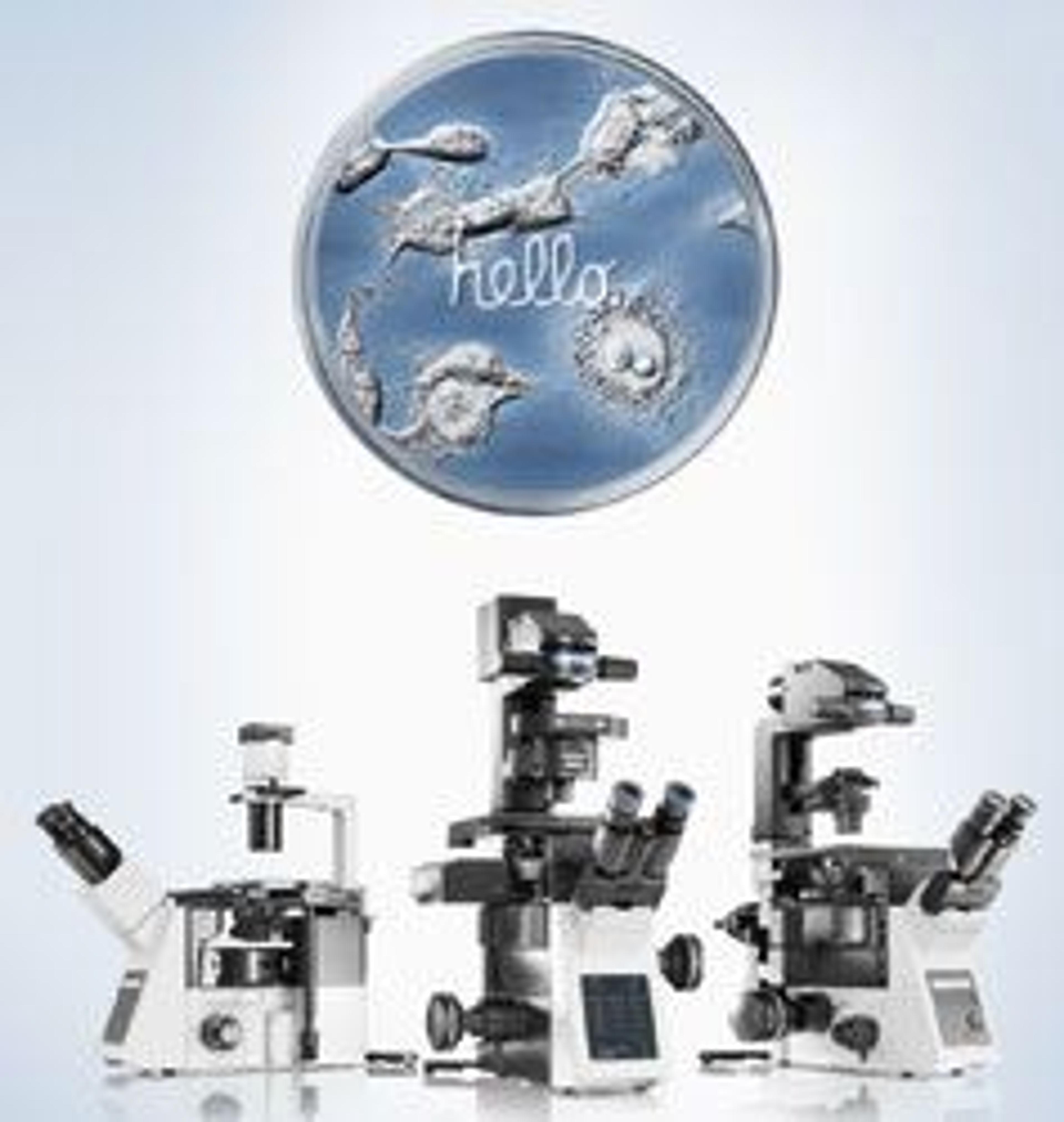
IX3 Series - Intuitive & open source Microscope Systems
EVIDENTThe series of flexible inverted microscope frames for live cell imaging
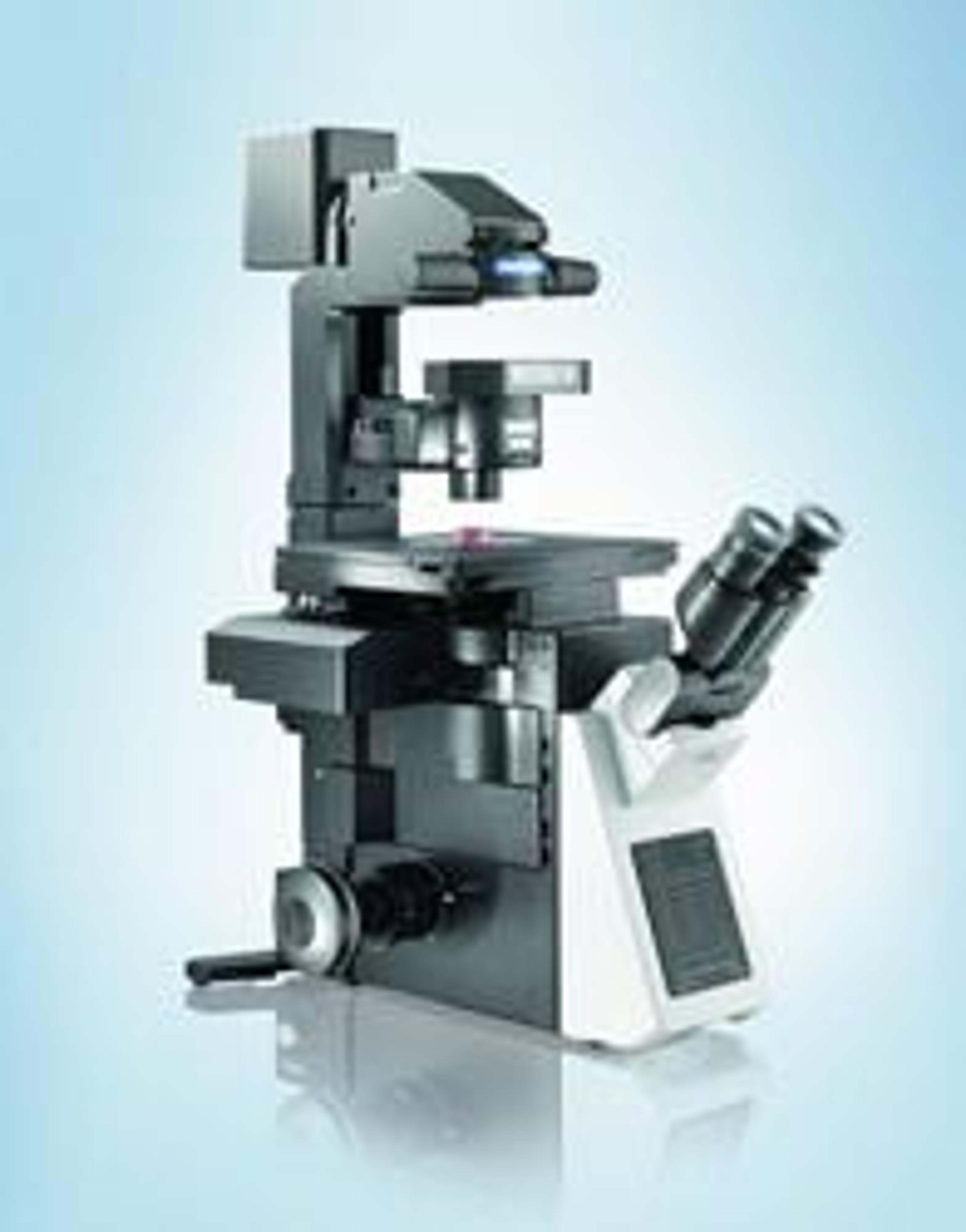
IX73 - Inverted Microscope System for Live Cell Imaging
EVIDENTExtended flexibility and performance in live cell imaging
IX83 - Fully-motorized and automated Inverted Microscope System
EVIDENTExtended flexibility and performance in live cell imaging