Olympus exhibits leading microscopy and inspection solutions at world’s first Virtual Analytical Summit

Olympus' virtual presence at the Summit will inform scientists around the globe about the latest techniques in microscopy and industrial inspection

24 Mar 2020
Diane Li
Assistant Editor

Industry news

Olympus will exhibit its leading microscopy and industrial inspection solutions to a global online audience at the new SelectScience Virtual Analytical Summit 2020, taking place from 31 March to 2 April 2020. The event is free to register and attend and will connect 900k+ scientists with presentations, workshops, video interviews, virtual booths, cutting-edge resources, the latest product and application news, Q&As, networking and live-chat opportunities.

As the spread of COVID-19 forces the postponement of key events in the scientific calendar, the Virtual Summit offers a crucial forum for scientists and leading manufacturers to continue to connect in order to advance science. The virtual summit will enable scientists and scientific manufacturers to come together from across the world to explore headline topics across many areas, including life science, pharma and materials science.

Responding to this need, Olympus will exhibit a wide range of solutions for applications ranging from routine cell culture through to high-content screening and elemental analysis for alloy identification, including:

  • Breakthrough high-performance X Line objectives for exceptional image quality for researchers and clinicians alike
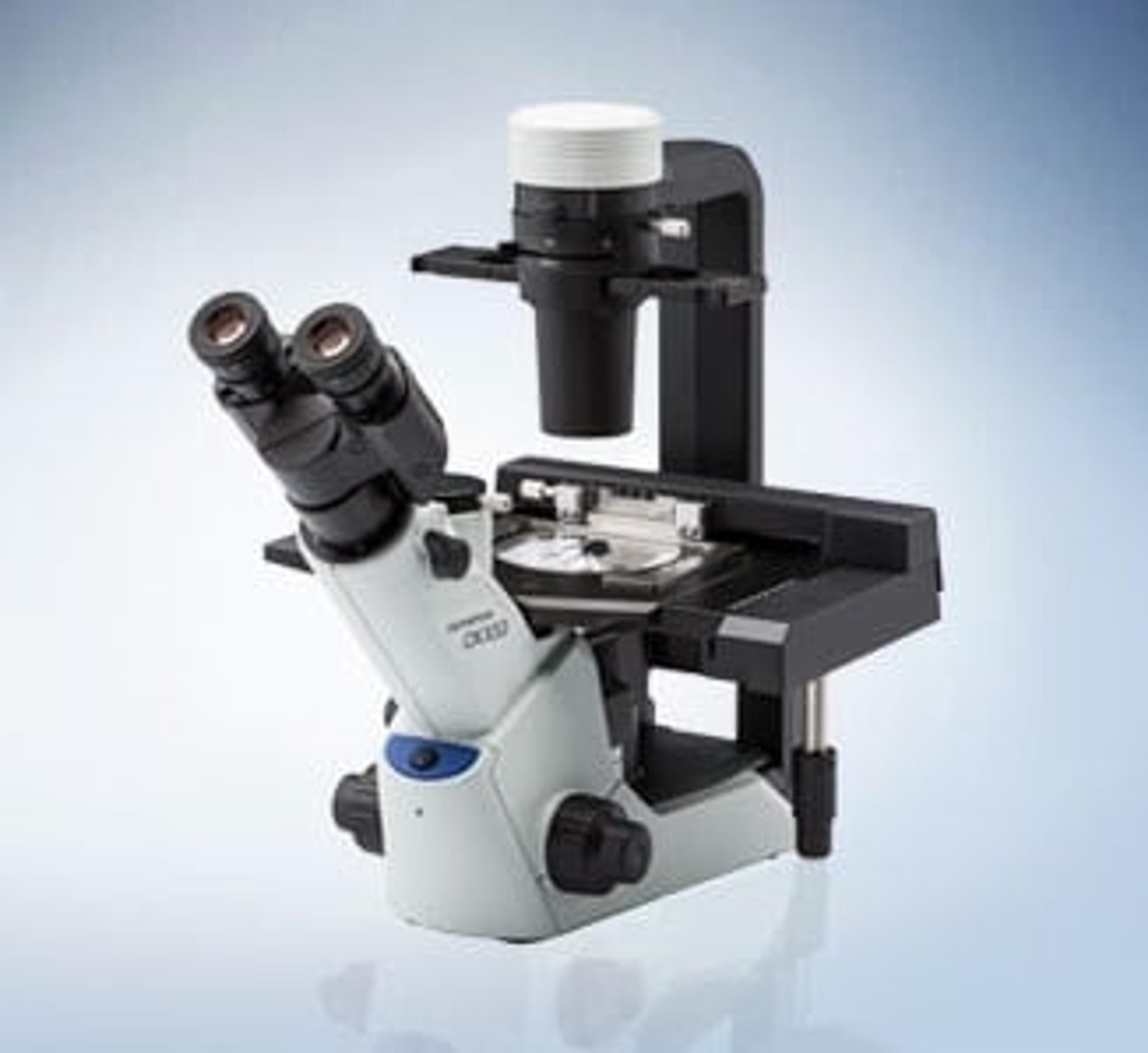
  • The compact, ergonomic CKX53 inverted cell culture microscope for routine wet lab use
  • The flexible, wireless EP50 color camera – perfect for use in classroom teaching
  • DSX1000 digital microscopes, which offer speed and versatility to materials inspection thanks to easy switching between six observation modes and a rotating head and stage
  • Vanta handheld XRF analyzers, which deliver rapid and accurate elemental analysis and alloy identification in the field
  • The SLIDEVIEW VS200 digital slide scanner for easy analysis, sharing and archiving of slides
  • The scanR high-content screening station featuring deep learning technology

Deep learning in microscopy has seen a rapid increase over the past years. To demonstrate the capabilities of the latest AI-based systems, Dr. Daniel Krüger of Olympus Soft Imaging Solutions will give a presentation at 2pm BST / 3pm CEST on 1 April, which will cover:

  • A practical introduction to deep learning and neural networks
  • Analysis examples of self-learning microscopy
  • Applications of deep learning in microscopy and high-content screening

The summit opens from 31 March to 2 April 2020 (11am–8pm BST / 12–9pm CEST) and delegates will be able to drop in and out as convenient.

Register for free to reserve a place today>>

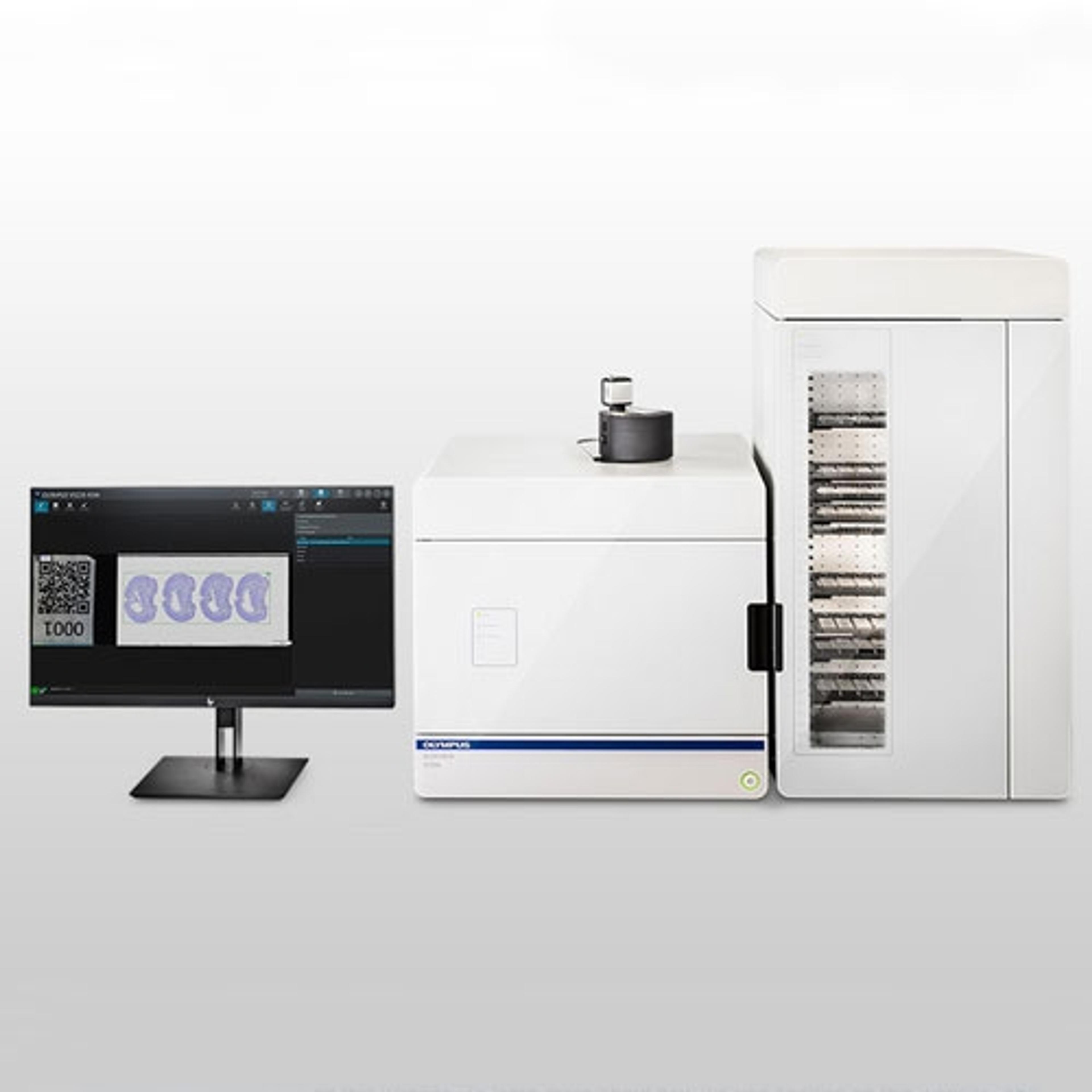
SLIDEVIEW VS200 Research Slide Scanner

EVIDENT

Easily analyze, share, and archive your data with the SLIDEVIEW VS200 digital slide scanner. Designed to capture high-resolution images of your slides for quantitative analysis, the system enables you to make the most of the information your slides have to offer.
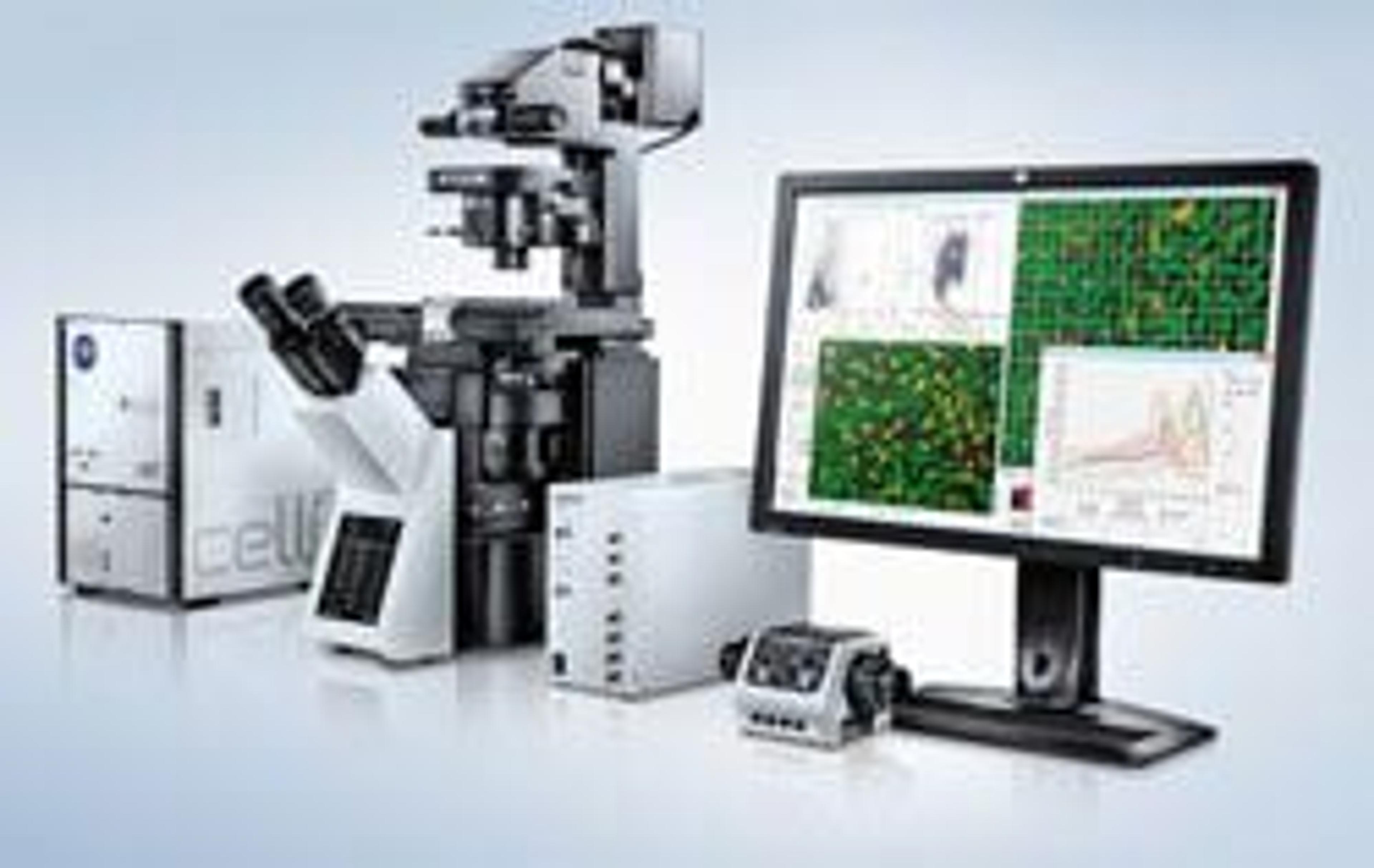
(0)

Links

Tags

In Vivo Imaging Systems<i>In vivo</i> imaging systems, including pre-clinical imaging systems and medical imaging systems are used to non-invasively visualize and capture images of live animals and plants. Monitor the natural processes or diseases of your subjects using small-animal pre-clinical imaging systems, including single photon positron emission tomography (SPECT), positron emission tomography (PET), computed tomography (micro-CT), magnetic resonance imaging (MRI), X-ray radiography, ultrasound, fluorescence and bioluminescence imagers. Multimodal systems and software solutions are also available for correlative analysis of organ, tissue, cell, or molecular-level processes. Find the best in vivo imaging products in our peer-reviewed product directory: compare products, check customer reviews and receive pricing direct from manufacturers.Light MicroscopyLight microscopes or optical microscopes are used to visualize microscale objects under magnification, including cells, clinical specimens and materials. Lab equipment for light microscopy includes confocal microscopes, fluorescence microscopes, zoom and stereo microscopes. Microscope slides and imaging reagents are available for visualizing samples, as well as various microscope stages and incubators for large or temperature-sensitive samples. Find the best light microscopes in our peer-reviewed product directory: compare products, check customer reviews and receive pricing direct from manufacturers.Electron MicroscopyElectron microscopes (EM) are used to create high-resolution images of samples at the nanoscale by means of an accelerated beam of electrons as a source of illumination. Types of electron microscope include scanning electron microscopes (SEM), transmission electron microscopes (TEM), scanning transmission electron microscopes (STEM) and cryo-electron microscopes. Focused ion beam (FIB) microscopes are useful for modifying or milling a sample surface with nanometer precision, as well as imaging. Find the best electron microscopes in our peer-reviewed product directory: compare products, check customer reviews and receive pricing direct from manufacturers.Digital MicroscopyDigital microscopy involves using digital cameras and sensors to capture high-resolution images of samples for analysis. It offers enhanced imaging capabilities compared to traditional optical microscopy and is widely used in biological and material science research. Explore digital microscopy systems in our peer-reviewed product directory; compare products, check reviews, and get pricing directly from manufacturers.CCD cameraFluorescence MicroscopyFluorescence microscopy has become an essential tool in biology, as well as in materials science. The application of many fluorochromes has made it possible to identify cells and sub-microscopic cellular components with a high degree of specificity. Using multiple fluorescence labels, different probes can simultaneously identify several target molecules.X-Ray FluorescenceX-ray fluorescence (XRF) is an analytical technique used to determine the elemental composition of materials. It is widely used in geology, materials science, and environmental testing for detecting heavy metals and other elements. Browse our peer-reviewed product directory to find the best XRF systems, compare products, check reviews, and get pricing directly from manufacturers.MicroscopyMicroscopy is a technique used to observe small objects in detail, from cells to materials, using light or electron microscopes. It enables researchers to examine structures with high resolution, aiding in fields such as biology, medicine, and materials science. With advanced microscopy techniques, scientists can gain insights into cellular processes, tissue structures, and material properties. Explore the best microscopy solutions in our peer-reviewed product directory, compare products, read customer reviews, and get pricing directly from manufacturers.Super Resolution Microscopy