Olympus at Analytica: Dedicated to Innovations in Microscopy
13 Mar 2014
Product news
Enabling a myriad of new and sometimes unusual applications throughout life and materials science, Olympus will demonstrate its aptitude for pushing the boundaries of light microscopy at Analytica, hall A2, booth 309.
In vivo Imaging: reaching new depths
Multiphoton microscopy presents a powerful tool within life science research, achieving fundamental insights into the intricate and complex workings of biological systems.
Olympus has responded to the need for a high-performance in vivo imaging system with the new FluoView FVMPE-RS. The system enables high-precision, ultra-fast scanning and stimulation – allowing researchers to see deep within specimens, take measurements at the highest speeds and capture images even when working under the most demanding conditions. In his presentation at the BIOTECH Forum (2nd April, 2pm), entitled “The new FluoView FVMPE-RS: an advanced multiphoton microscope for imaging with high speed and extended IR range”, Olympus application specialist Dr Bjoern Sieberer will explain how these capabilities advance a range of in vivo imaging applications.
Dedicated to life science – build your own microscope
Enhancing live cell imaging applications, the IX3 introduces ultimate flexibility with ‘open source’ microscopy, and visitors to the booth can experience first-hand how bespoke systems are effortlessly created by exchanging optical modules from an accessible infinite light path.
Centered on workflow ergonomics with a modular and flexible design, the cellVivo incubation system presents the ideal complement to the open source frame, and visitors will be able to see just how simple it really is to expand the capabilities of the high-end IX83 for advanced live cell imaging. As well as precise control of environmental conditions and user-friendly remote monitoring, cellVivo is uniquely accessible with just one hand, enabling simultaneous handling of the incubator and specimens for swift usage, protecting samples from prolonged exposure to the external environment.
Bringing operating simplicity to materials science
Enhancing the efficiency of non-destructive inspection and metrology, the Olympus opto-digital range will also be on show, including the award-winning DSX and the new LEXT OLS4100.
With precision optics and DIC capability enabling resolving power beyond that of a typical light microscope, the LEXT has found an unusual utility within the area of life science. Swiss researchers working in reproductive toxicology have used the LEXT for the morphological analysis of sperm, and will be at the booth to discuss how the LEXT has enhanced their work with its potential to be used in place of scanning electron microscopy.
To find out more about Olympus microscope systems and their uses, visit hall A2, booth 309.
www.olympus-europa.com/microscopy
www.olympus-ims.com/en/microscope
Related products
Request Quote for All Products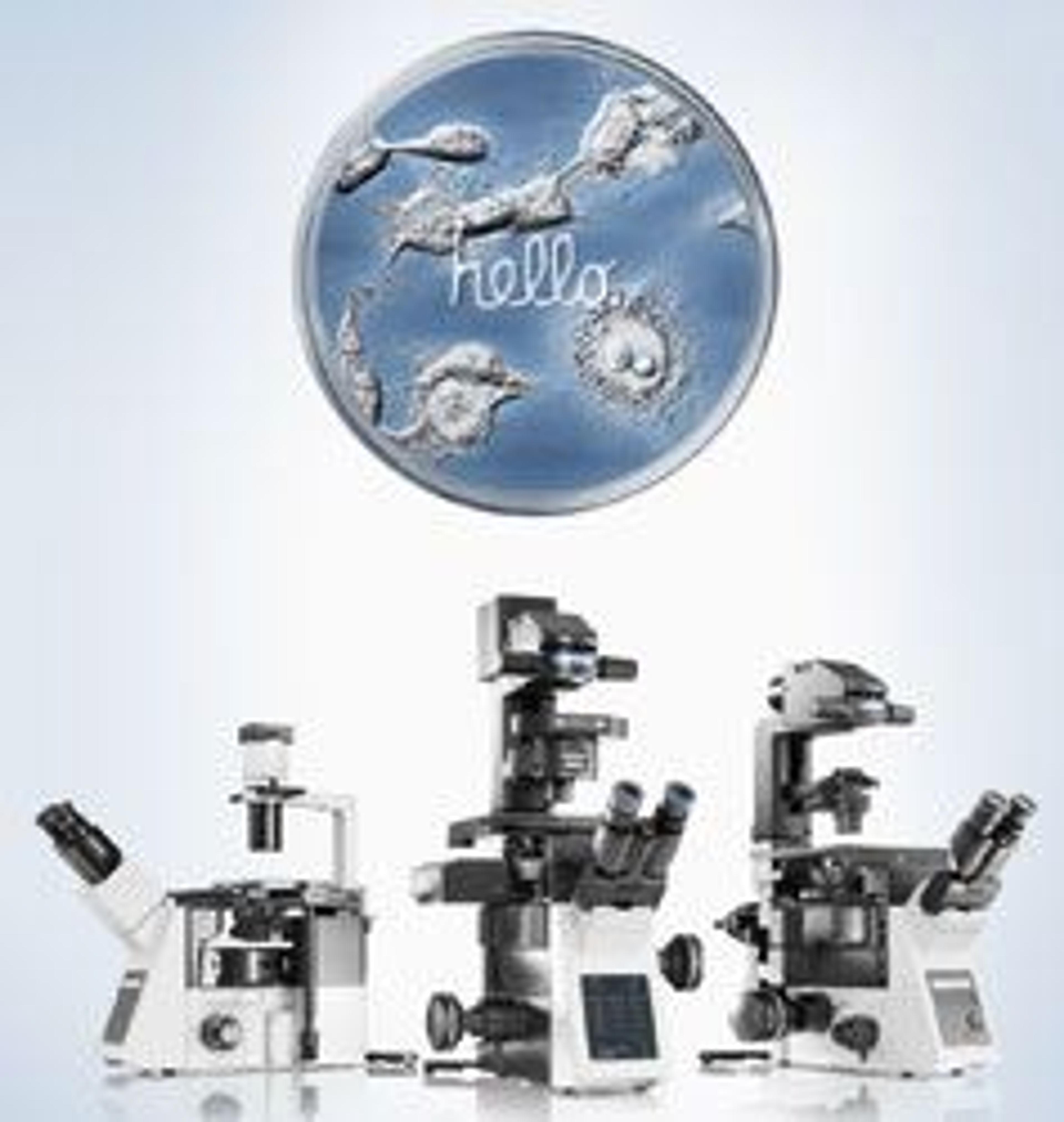
IX3 Series - Intuitive & open source Microscope Systems
EVIDENTThe series of flexible inverted microscope frames for live cell imaging