Novel In-vivo Imaging Solution for Neuroscientists

4 Apr 2016
Alex Waite
Editorial Assistant

Product news

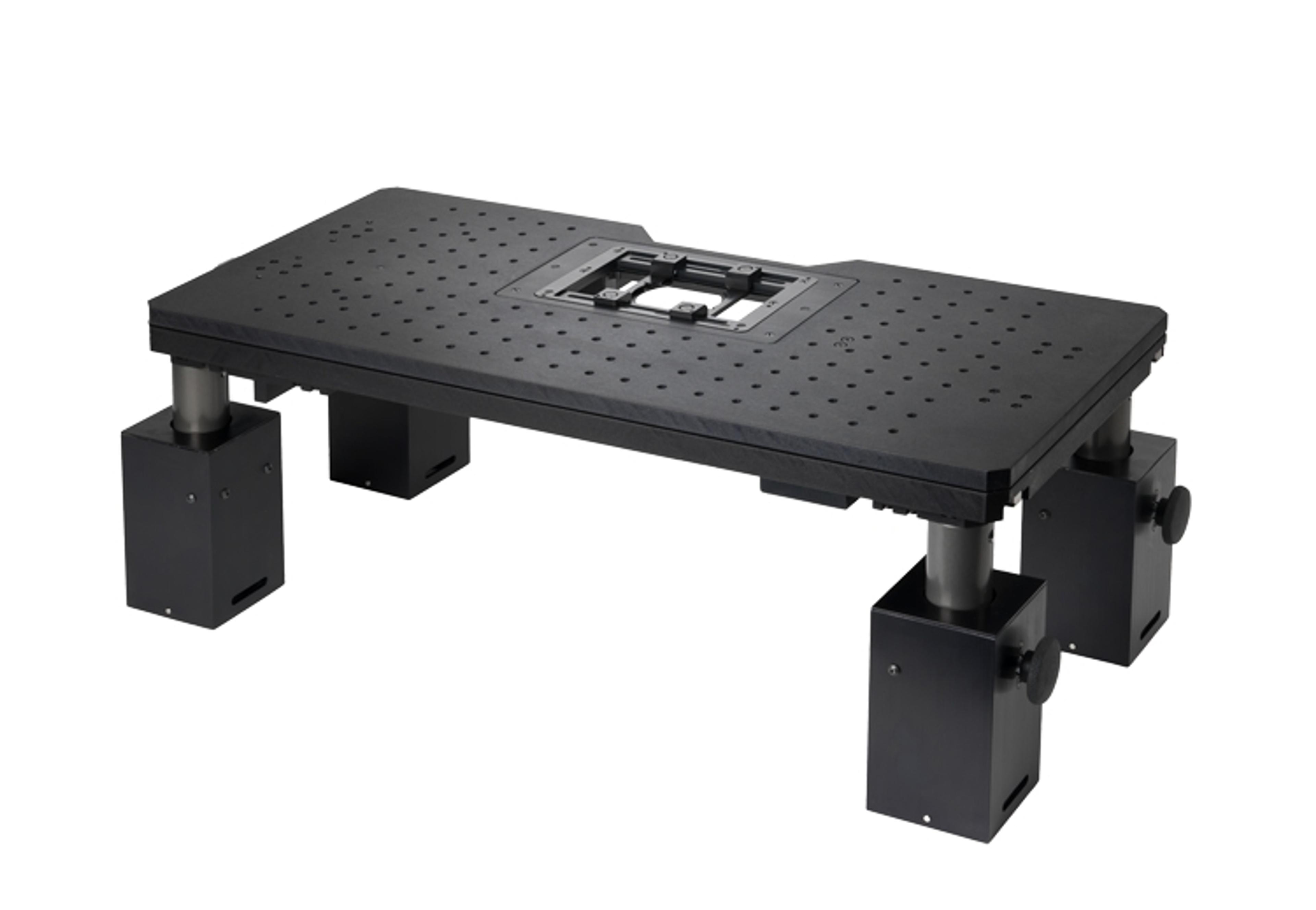
Prior Scientific reports that its collaboration with Neurotar (Helsinki, Finland) has produced a seamlessly integrated solution for in vivo microscopic imaging in the brain of awake and moving rodents. The solution is based upon Prior’s ultra-stable and easily adjustable Z-Deck platform.

Neurotar’s Mobile HomeCage™ is an accessory device for microscopy and electrophysiology, which enables high precision tests in the brain of awake, head-fixed, but otherwise freely moving rodents. The Mobile HomeCage eliminates the need for anesthesia and thus preserves full physiological functioning of the brain. It provides a natural, tangible environment, which alleviates the stress experienced by rodents during experiments under standard head-fixation conditions. Stress reduction helps reduce the run-up time to the start of experiments and improves their reproducibility. The Mobile HomeCage is compact – it fits into most imaging set-ups without the need for modifications. Last but not least, it allows combining state-of-the-art neurophysiological techniques (such as in vivo two-photon microscopy, optogenetics, intrinsic optical imaging and in vivo patch-clamp) with behavioral paradigms in a single experiment.

Working closely with Neurotar, Prior have developed a fixing kit allowing seamless integration of the Mobile HomeCage with the Z-Deck. The Z-Deck is an ultra-stable, height adjustable platform designed specifically for electrophysiology and microscopy applications in neuroscience, which is available with an integral motorized positioning stage, a manual positioning stage, or a fixed top plate. The Z-Deck is compatible with a wide range of microscopes.

Available from Neurotar (www.neurotar.com) the new fixing kit allows neuroscientists to combine the benefits offered by the Mobile HomeCage with the stability, height adjustment and operational flexibility offered by the Z-Deck. Importantly, the fixing kit allows the loading of the mouse either on or off the stage followed by a quick and precise re-positioning of the Mobile HomeCage onto the Z-Deck. This means that the delicate operation of mouse-loading does not need to be performed around the microscope system itself.

Links

Tags

Sample PreparationSample preparation can improve the quality and speed of separation techniques. Products to assist sample preparation include filtration equipment, evaporators, membranes and sieves.Data AnalysisData analysis hardware and software is available to make data processing straight-forward yet powerful. Data software can be used for math and stats, technical graphing and image analysis. In addition, software is available for specific data analysis of electrophoresis, densitometry, ELISA and DNA sequencing.Sample ManagementSample management systems include sample storage devices such as freezers and plate storers, sample environment enclosures and sample organization, retrieval and sorter systems. Useful system features include high-throughput, automation, robotic arms, automated liquid handling and associated database systems. Accessories in sample management include barcode scanners, heat sealers and tubes.Cellular PathologyCellular Pathology deals with the microscopic analysis of tissue samples and cells. Sample preparation and processing includes fixation, staining, sectioning and slide mounting, using equipment such microtomes and cryostats. In choosing immunohistochemistry and immunocytochemistry kits, consider chromogens, staining method, antibodies, microscopes and imaging.In Vivo Imaging Systems<i>In vivo</i> imaging systems, including pre-clinical imaging systems and medical imaging systems are used to non-invasively visualize and capture images of live animals and plants. Monitor the natural processes or diseases of your subjects using small-animal pre-clinical imaging systems, including single photon positron emission tomography (SPECT), positron emission tomography (PET), computed tomography (micro-CT), magnetic resonance imaging (MRI), X-ray radiography, ultrasound, fluorescence and bioluminescence imagers. Multimodal systems and software solutions are also available for correlative analysis of organ, tissue, cell, or molecular-level processes. Find the best in vivo imaging products in our peer-reviewed product directory: compare products, check customer reviews and receive pricing direct from manufacturers.ElectrophysiologyElectrophysiology is the study of biological voltage changes, from a single ion channel through to a whole organ scale. Voltages can be measured using patch clamping, automated workstations, electrophysiology rigs or cell based assays. Specific cell lines, manipulators and microinjectors are also available for electrophysiology research.Light MicroscopyLight microscopes or optical microscopes are used to visualize microscale objects under magnification, including cells, clinical specimens and materials. Lab equipment for light microscopy includes confocal microscopes, fluorescence microscopes, zoom and stereo microscopes. Microscope slides and imaging reagents are available for visualizing samples, as well as various microscope stages and incubators for large or temperature-sensitive samples. Find the best light microscopes in our peer-reviewed product directory: compare products, check customer reviews and receive pricing direct from manufacturers.Animal ModelsThe use of non-human animals in experiments or behavorial observations. The research is conducted inside universities, medical schools, pharmaceutical companies, farms, defence establishments, and commercial facilities that provide animal-testing services to industry. It includes pure research such as genetics, developmental biology, behavioral studies, as well as applied research such as pharmaceutical testing in pre-clinical, before human, studies. RegulatoryPharmaceutical regulations impact on all areas of drug development, manufacture and control and supply. Services are provided from the early stages of drug development and clinical trials through to dossier submission, approval and marketing. Regulatory also includes submission of analytical studies from the pharmaceutical product and safety. NeuroscienceNeuroscience research investigates the neurological mechanisms underlying behavior, neurodegenerative diseases, and other brain conditions. Learn about the innovative technologies for bioimaging, electrophysiology, cell culture, chromatography and other techniques used in this field.NeurobiologyNeurobiology studies the biology of the nervous system, including the structure and function of neurons, glial cells, and their interactions. It is a key field in understanding neurological disorders such as Alzheimer�s, Parkinson�s, and schizophrenia. Research in neurobiology is essential for developing therapies for these conditions. Browse our peer-reviewed product directory to find the best neurobiology research tools, compare products, check reviews, and get pricing directly from manufacturers.Small Animal ImagingSmall animal imaging is the visualization of living animals, most commonly mice and rats, for research purposes. This non-invasive technique is vital for long-term studies into drugs effects, as well as small animal anatomy and physiology.Optical Gas ImagingMicroscopyMicroscopy is a technique used to observe small objects in detail, from cells to materials, using light or electron microscopes. It enables researchers to examine structures with high resolution, aiding in fields such as biology, medicine, and materials science. With advanced microscopy techniques, scientists can gain insights into cellular processes, tissue structures, and material properties. Explore the best microscopy solutions in our peer-reviewed product directory, compare products, read customer reviews, and get pricing directly from manufacturers.
Novel In-vivo Imaging Solution for Neuroscientists