NEW Generation Syngene Imaging Systems for Gel and Blot Imaging
The new systems will be live on Stand 140 at ARABLAB 2017
25 Jan 2017
Product news
Syngene, a world-leading manufacturer of image analysis solutions, is delighted to announce the new range of imaging systems will be on Stand 140 at ARABLAB 2017 on March 20th-23rd. Designed for simple set-up and accurate performance, there will be systems on display to suit all budgets and imaging applications.
Scientists looking for a low cost, yet powerful DNA and protein imaging system, should check out Syngene’s NuGenius, live on stand. This compact gel doc system, featuring a 5 million pixel CCD camera and motor driven zoom lens, enables perfect imaging of any gel or blot size up to 20cm x 24cm.
For those seeking dedicated chemiluminescence imagers, Syngene’s experts will be on hand to show the brilliantly designed GeneGnome XRQ imager. With an innovative low-light capture powerhouse of a camera and one-click set-up, generating superb images of barely-there chemiluminescent bands on Western blots has never been easier.
To complete Syngene’s range, the PXi all-round multi-functional imaging system is available for scientists to try. This system, complete with high resolution, cooled camera and innovative touch-screen GeneSys image capture software can handle anything with ease, from chemiluminescent and fluorescent blots, visible gels and blots, right through to more complex 2D gels.
“ARABLAB is always such a great show and we’re excited to be exhibiting our latest range of intelligently designed gel doc systems here.” comments Matthew Dunne, Senior Divisional Manager at Syngene. “We look forward to welcoming scientists on Stand 140 and to letting them see how imaging technology produced in one of the world’s most renowned innovation hubs, could significantly benefit their research.”
Related products
Request Quote for All Products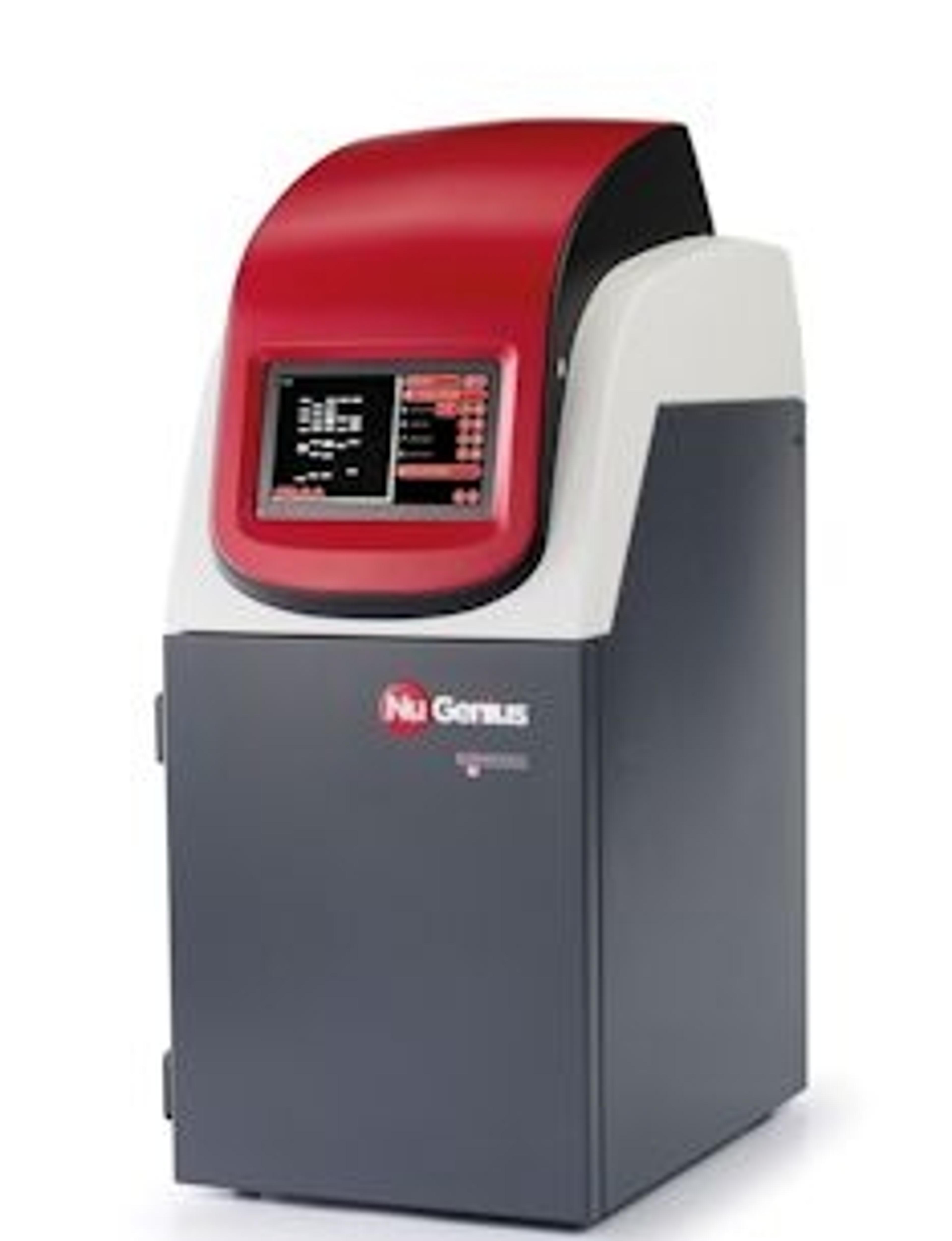
GeneSys
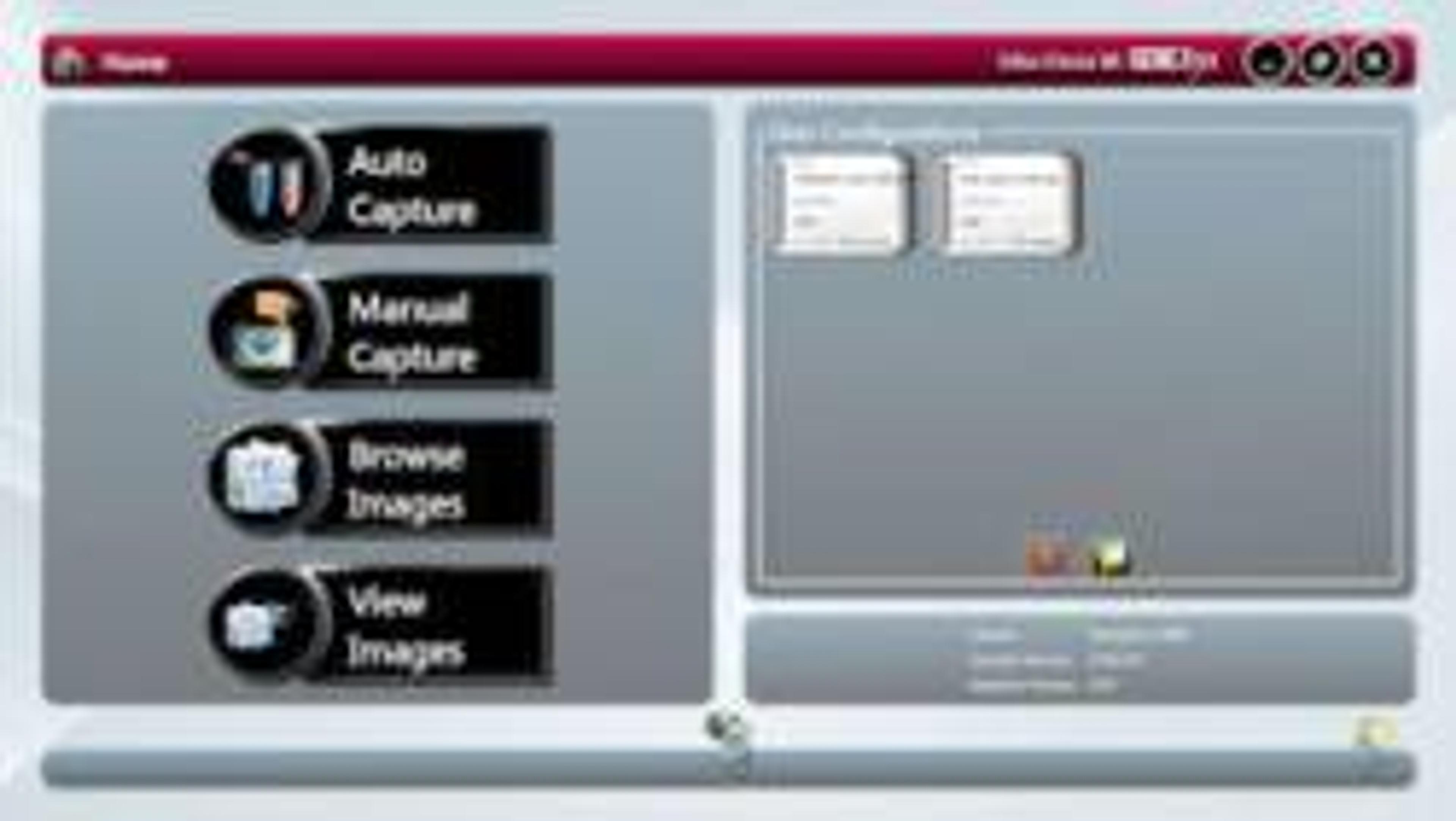
SyngeneThe biggest difference between a Syngene system and any other is in the way the system is controlled. At the heart of all Syngene systems is GeneSys which is an application driven control software. You can use GeneSys in either Automatic or Manual mode. It is assumed that the user will know exactly what their application is and how they have prepared their gel or blot. In Auto Mode they simply enter this information into GeneSys (or recall it from a saved configuration) and the system takes over the rest. Behind GeneSys is an extensive database which contains data relating to a very wide range of applications, eg, fluorescence, chemiluminescene and chemifluorescence.Once the imaging system has been told what sample to expect then GeneSys decides what hardware configuration is best and sets the system ready for image capture. Things like camera control, exposure time, sensitivity setting, lighting requirements, lens control, filter selection – all of this is taken care of by GeneSys. The user just has to click the ‘capture’ button and wait for the perfect image to appear. GeneSys ensures scientists can quickly capture excellent images of even complex multiplex gels.The innovative GeneSys software features large touch-screen buttons which guide users effortlessly through set-up and image capture. Each screen prompts researchers to select, for example, the type of gel or blot they are using and what it is stained with.

GeneGnome XRQ
SyngeneGeneGnome is a dedicated chemiluminescence imaging system. Utilising the GeneGnome XRQ’s optimised short ‘camera to sample’ technology, you’ll get more than double the dynamic range of film, making it easy for you to see picogram or femtogram protein levels without all the fuss.