NemaMetrix Introduces Fluorescent Staining Kits for Improved Phenotyping in model organism C. elegans

7 Jun 2017
Lois Manton-O'Byrne, PhD
Executive Editor

Product news

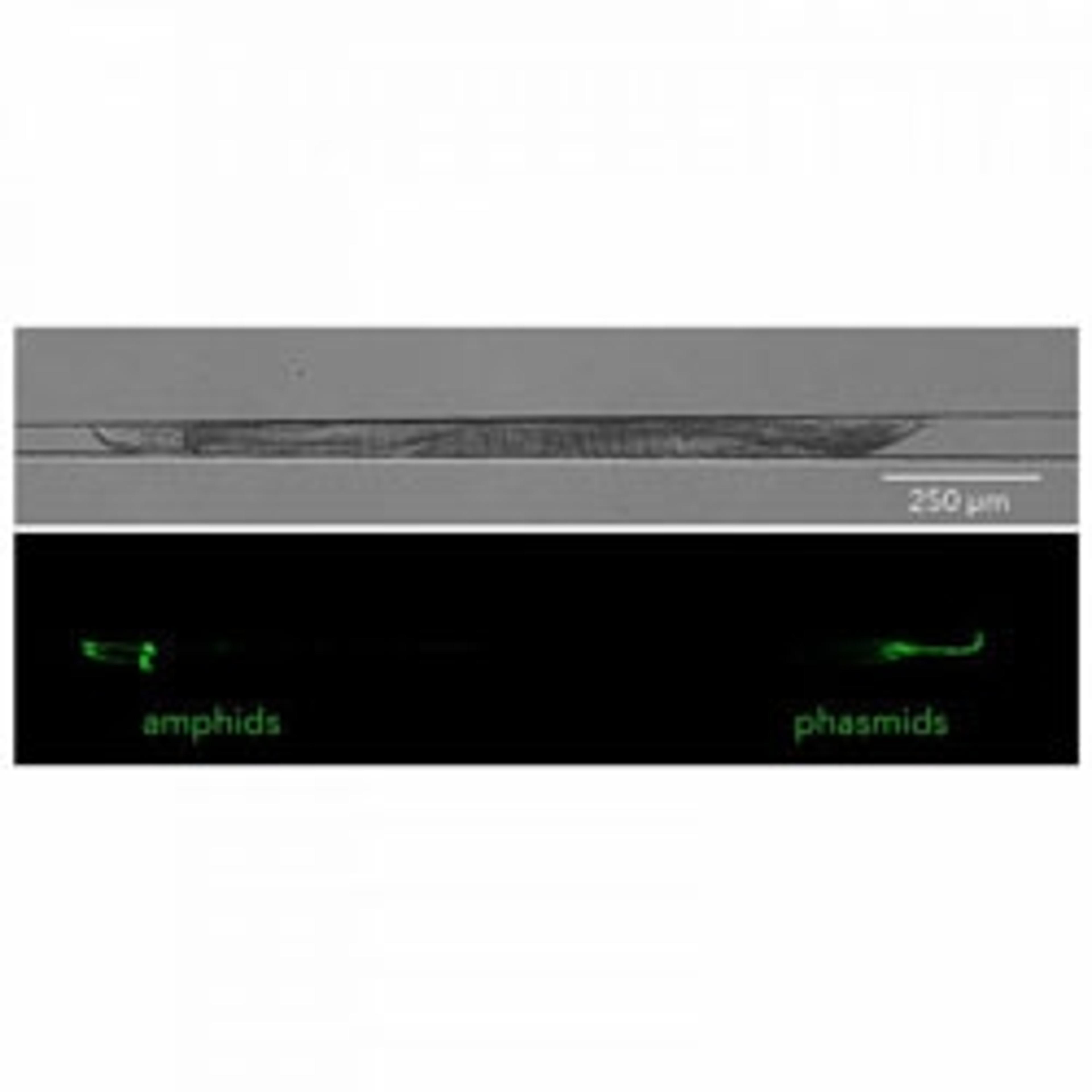
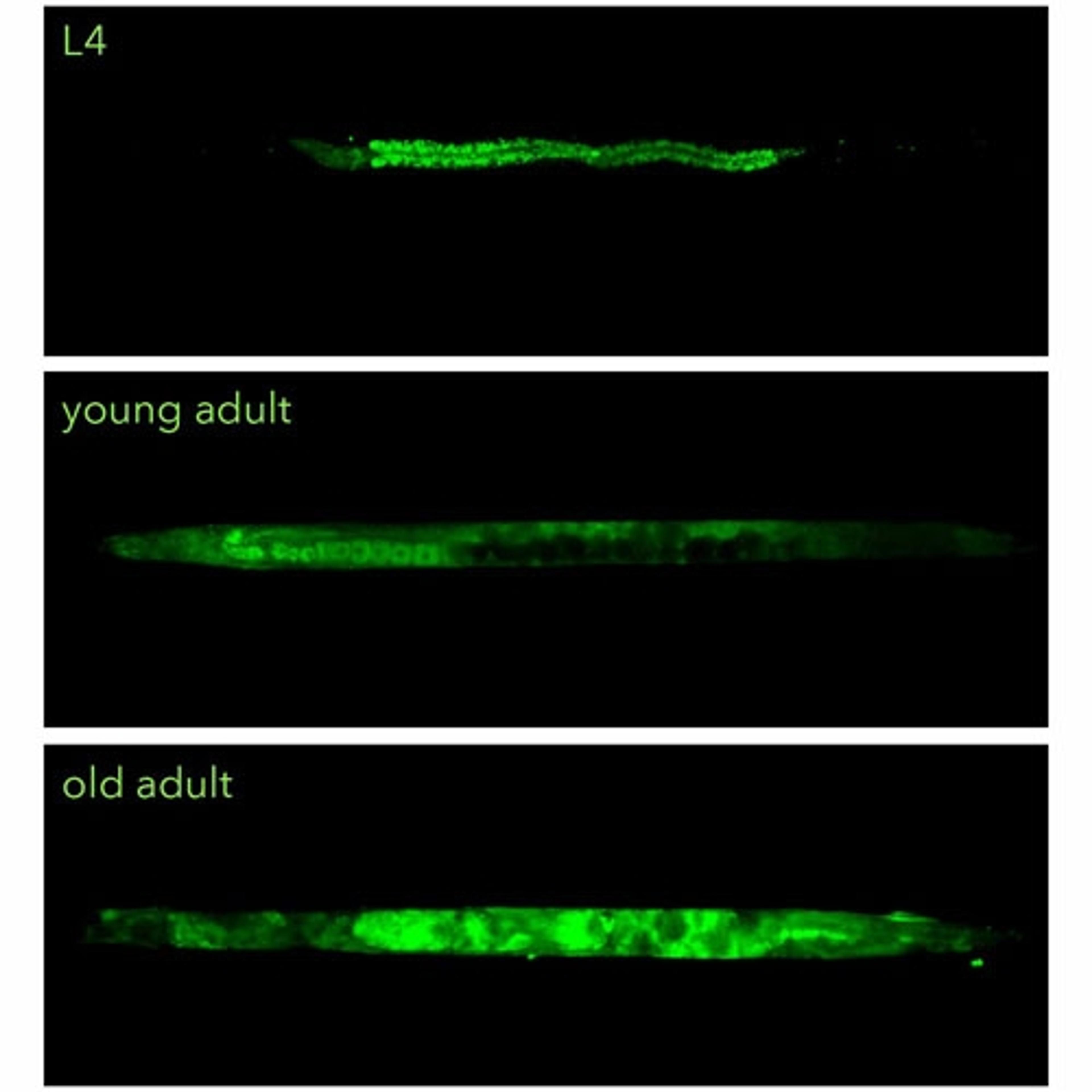
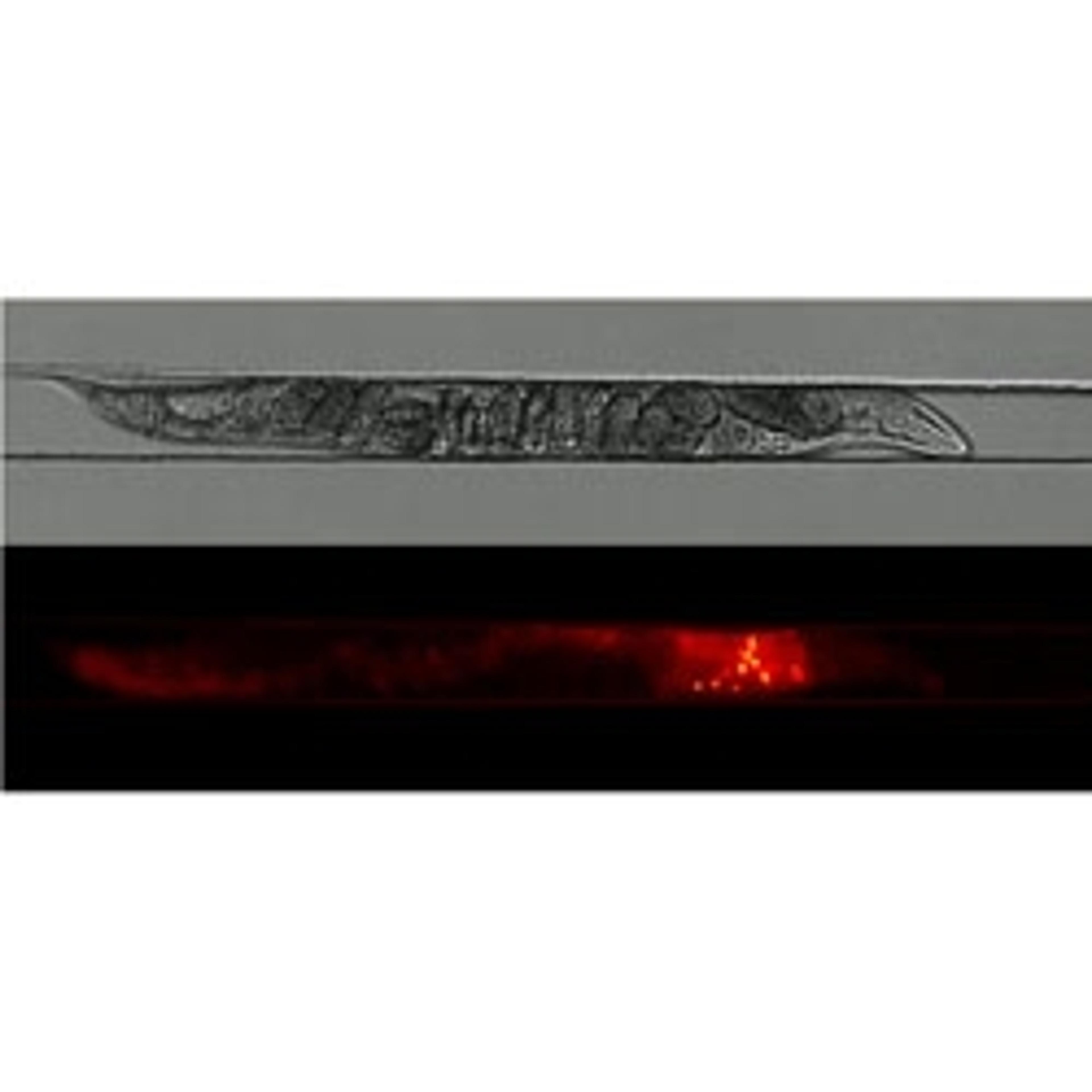
NemaMetrix, a company developing nematode-based drug development and screening technologies, today announced the launch of its first series of RediStain™ fluorescent kits for improved visualization of C. elegans’ structure and function in phenotyping studies. These kits expand the toolkit of C. elegansresearch to include the ability to perform structural and functional assays that are standard in cell biology.

Used with the company’s ScreenChip System, the new staining kits provide a more efficient way to study nematode phenotypes and their directly-related underlying genotypes, which would not be possible with non-fluorescent techniques.

NemaMetrix has developed the ScreenChip System, a first-in-class automatic screening and phenotyping platform utilizing C. elegans as the model organism, which acquires and displays neuromuscular data in real time. The technology allows researchers to rapidly generate and collect precise whole-animal data via quick and reliable assessment of neuronal and physiological responses to genetic mutations, drug effects and other environmental changes. The platform provides an alternative to enable researchers to better understand human diseases and explore potential treatments for high-impact disorders. The ScreenChip System is currently being used in aging, neurodegenerative, and cardiac diseases studies, as well as in drug development and developmental biology.

C. elegans is one of the world’s most well understood model organisms; the nematode worm has phenotypes that can be directly related to human genes, with homologues identified for up to 80% of human genes, and a lifespan of just 14 days. This makes it a powerful experimental model for investigating human disease, allowing researchers to explore and discover new phenotypes that are difficult and expensive to assess using traditional models and methods.

Matt Beaudet, CEO of NemaMetrix commented: “C. elegans is an inexpensive, live-animal model which has the potential to revolutionize the drug development process. The introduction of fluorescent staining kits to our ScreenChip System will make previously invisible phenotypes easy to visualize, allowing scientists to generate more and better quality data on neuronal and physiological responses to a wide range of genetic and environmental changes.”

Links

Tags

Gene Expression and Molecular CloningMolecular cloning is a set of techniques that utilizes vectors to transfer recombinant DNA into host cells and is an essential tool for investigating the expression of genes and proteins in bacterial or mammalian cells. A variety of vectors optimized for gene cloning and expression in a range of host organisms are available, alongside competent cells for genetic replication. Here, you can explore a range of molecular tools, high-quality genomic and cDNA libraries, premade clones, transformation and transfection reagents and mutagenesis or gene expression detection assays and expression arrays. Find the best gene expression and molecular cloning products in our peer-reviewed product directory: compare products, check customer reviews and receive pricing direct from manufacturers.Biopharmaceutical AdvancesBiopharmaceutical advances follow the development of pharmaceuticals derived from biotechnology, also known as biotechnology medicines. Biopharmaceuticals may be produced from cell lines, plants, or microbial cells. Important considerations of biopharmaceutical use include application, cost, production process and purification.In Vivo Imaging Systems<i>In vivo</i> imaging systems, including pre-clinical imaging systems and medical imaging systems are used to non-invasively visualize and capture images of live animals and plants. Monitor the natural processes or diseases of your subjects using small-animal pre-clinical imaging systems, including single photon positron emission tomography (SPECT), positron emission tomography (PET), computed tomography (micro-CT), magnetic resonance imaging (MRI), X-ray radiography, ultrasound, fluorescence and bioluminescence imagers. Multimodal systems and software solutions are also available for correlative analysis of organ, tissue, cell, or molecular-level processes. Find the best in vivo imaging products in our peer-reviewed product directory: compare products, check customer reviews and receive pricing direct from manufacturers.Light MicroscopyLight microscopes or optical microscopes are used to visualize microscale objects under magnification, including cells, clinical specimens and materials. Lab equipment for light microscopy includes confocal microscopes, fluorescence microscopes, zoom and stereo microscopes. Microscope slides and imaging reagents are available for visualizing samples, as well as various microscope stages and incubators for large or temperature-sensitive samples. Find the best light microscopes in our peer-reviewed product directory: compare products, check customer reviews and receive pricing direct from manufacturers.Animal ModelsThe use of non-human animals in experiments or behavorial observations. The research is conducted inside universities, medical schools, pharmaceutical companies, farms, defence establishments, and commercial facilities that provide animal-testing services to industry. It includes pure research such as genetics, developmental biology, behavioral studies, as well as applied research such as pharmaceutical testing in pre-clinical, before human, studies. Fluorescence MicroscopyFluorescence microscopy has become an essential tool in biology, as well as in materials science. The application of many fluorochromes has made it possible to identify cells and sub-microscopic cellular components with a high degree of specificity. Using multiple fluorescence labels, different probes can simultaneously identify several target molecules.Small Animal ImagingSmall animal imaging is the visualization of living animals, most commonly mice and rats, for research purposes. This non-invasive technique is vital for long-term studies into drugs effects, as well as small animal anatomy and physiology.Phenotypic ScreeningPhenotypic screening assesses cellular responses to compounds, enabling drug discovery and target identification. This technique is pivotal in finding effective treatments for complex diseases. Discover phenotypic screening platforms and tools with peer-reviewed comparisons and pricing in our directory.Nematode Worms