Molsid Launch New "In Vivo GPS"
25 Sept 2018
Product news

An innovative family of pro-fluorescent, enzyme-responsive probes are introduced which act as an in vivo GPS. After 15 years of research at the Ecole Normale Supérieure de Lyon (ENS Lyon), Molsid S.A.S. launch these fluorogenic reagents for the rapid detection and accurate localization of selected enzyme activities in live cells. Their unique property is the release of a stable precipitating fluorochrome; this paves the way for significant technological improvements in areas such as medical diagnostics, quality control in the food industry, and industrial microbe development for the benefit of base chemical and pharmaceutical production as well as environmental decontamination.
Breakthrough innovation
The direct or indirect detection of an enzyme is at the heart of many Life Science processes. Since the 1970s, chemists and biologists have been seeking to develop synthetic fluorescent substrates that can unambiguously locate enzyme activities in a live cell. Up to date, the diffusion of the fluorescent signal in the sample remains the main defect of commercially available pro-fluorescent, enzyme-responsive probes, explaining their low sensitivity of detection.
A unique and patented chemical design allows Molsid to manufacture enzyme substrates with the following key features:
- Initial substrates are non-fluorescent, soluble molecules showing good cell permeability and long-term stability
- The product of probe conversion by the target enzyme is a bright, extremely photostable and crystalline fluorophore ensuring exceptional detection sensitivity
- Full signal retention at the site of enzymatic activity
- Sharp response
- Easy-to-use protocol without the need for secondary reagents
- End-to-end compatibility under physiological conditions
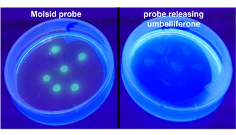
"Our precipitating probes make visible what standard probes do not allow to visualize. They provide clear and reliable results, thanks to a retention of the fluorescent signal at the seat of the targeted enzyme activity " explains Jens Hasserodt, Professor of Chemistry at ENS Lyon, inventor of the technology and co-founder of the company.
Bring new technical solutions to the market
The new range of fluorescent substrates, specific for several classes of hydrolases (glycosidases, peptidases and esterases/lipases) offers new solutions for researchers and engineers to set new standards for many high-value technologies: rapid microbiological analysis, detection of antibiotic resistance, diagnosis of biomarkers, medical imaging and enzyme engineering within host microbes for food and
green industries.