MILabs Launches New Molecular Imaging Product Line at the European Association of Nuclear Medicine Annual Meeting
20 Oct 2014
Product news
MILabs BV has launched a complete new preclinical product line at the 2014 Annual European Association of Nuclear Medicine Meeting in Gothenburg, Sweden, October 18-22. It consists of the new 4-series comprising U-SPECT4, U-SPECT4CT, VECTor4 and VECTor4CT incorporating significant improvements over the former PLUS-series. The 4-series carries even further the tradition of MILabs to deliver the best SPECT and PET image resolution available in the market.
The new systems are more compact and include a dramatically improved X-Ray CT subsystem with reduced radiation dose, increased resolution and speed. The CT subsystem comes at three different resolution levels. The high resolution versions have significant added value for application areas such as in vivo bone, cancer and arthritis research.
“Our customers are highly enthusiastic about the integrated low dose CT units and we are pleased to offer them dramatically improved resolution as well“, says Frederik Beekman, CEO/CSO of MILabs. This new generation U-SPECT4CT and VECTor4CT brings MILabs even further ahead in terms of combined ultra-high resolution functional and anatomical nuclear imaging. MILabs expects that the next generation U-SPECT4, U-SPECT4CT, VECTor4 and VECTor4CT systems will further accelerate the rapid growth of MILabs’ global customer base.
Related products
Request Quote for All Products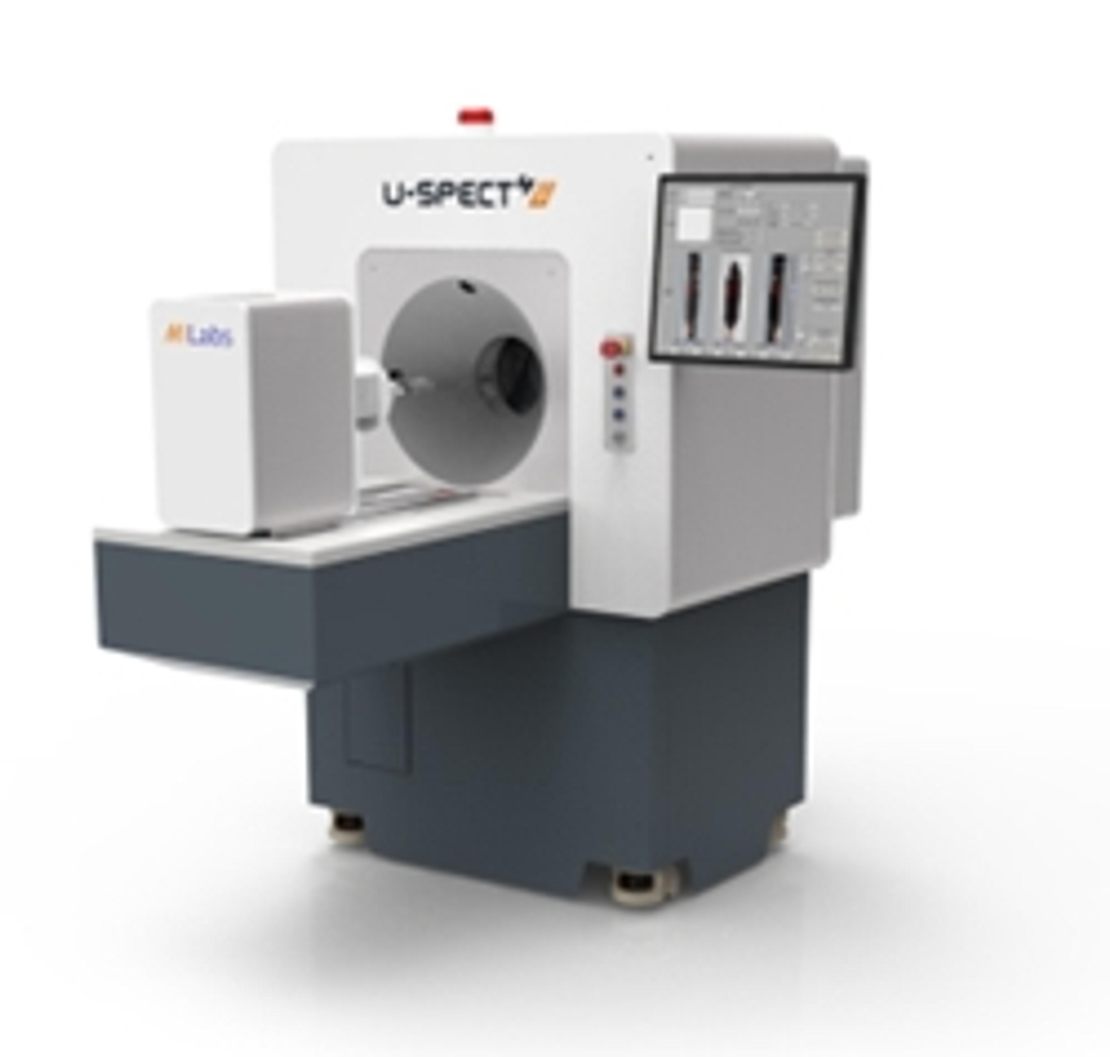
U-SPECT4 CT
MILabs BVBy far the best available SPECT resolution and sensitivity on the market. Patented 3D focusing capability for dynamic nuclear microscopy in organs and tumors. Ultra-fast imaging with multi-physiologic gating reveals dynamic processes with unprecedented time resolution, ideal for pharmacokinetic imaging.Patented M5™ collimation satisfies widest range of resolution, sensitivity and FOV needs. High reliability and long life due to very robust stationary detector design. Highly quantitative single and multi-tracer SPECT. Multi-modal imaging support for third party devices.Features: ¼ mm resolution Ultra-fast & sensitive (>13000 cps/MBq) Optional fully integrated CT