NewsSpectroscopy
lumox® and x-well Cell Imaging Consumables from SARSTEDT
3 Sept 2015
Lois Manton-O'Byrne, PhD
Executive Editor
Product news
Regardless of whether you are carrying out fluorescence- or light microscopic analyses on living or fixated cells, individual analyses or parallel test series, the comprehensive lumox® and x-well product range from SARSTEDT provides ideal solutions for superior optical results of your cell imaging tests.
x-well Cell Culture Chambers:
- x-well cell culture chambers are available with slides made of PCA, glass, cover glass or the lumox® film with excellent optical properties for optimal results in your cell imaging tests.
- The high-quality slide surfaces are ideally suited for the cultivation of adherent cells.
- All steps of histological and fluorescence staining can be conveniently carried out in a time-efficient manner.
- Small compartments enable most efficient testing by reducing cell numbers and reagents.
- Easy and convenient handling – The chambers of all products marked ”detachable“ can be detached from the slide without a tool, leaving no adhesive residues on the slide.
lumox® multiwell &dish
- The gas-permeable, highly transparent film base is ideal for excellent results in microscopic analyses of living and fixated cells.
- Black 24-, 96- and 384-well cell culture plates with lumox® film base (50 µm) for fluorescence microscopy.
- lumox® dishes (25 µm film base) are available with a diameter of 35 mm and 50 mm.
- For further analyses, (e.g. electron microscopy) the lumox® film can be excised.
Related products
Request Quote for All Products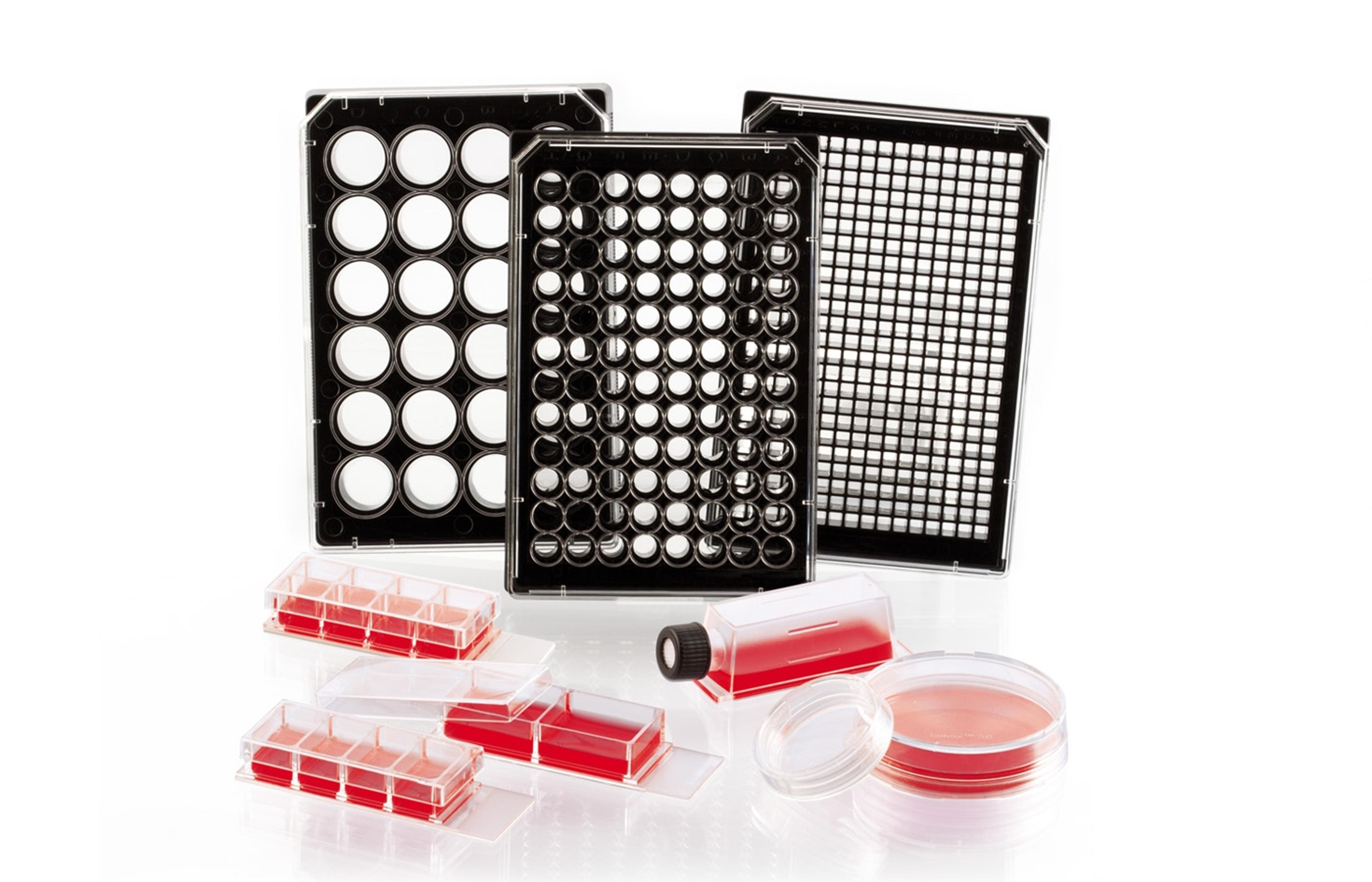

Links
Tags
Fluorescence SpectroscopyFluorometers and spectrofluorometers (also called fluorescence spectrometers) are used to measure the intensity and wavelength of fluorescent light emitted from a sample after excitation by illumination. Spectrofluorometers utilize monochromators to select the desired wavelengths, whereas filter fluorometers employ a set of filters. Spectrofluorometers for measuring steady-state fluorescence and lifetime fluorescence (or time-resolved fluorescence) are available, as well as fluorescence microscopes and microplate readers. Find the best fluorescence spectroscopy products in our peer-reviewed product directory: compare products, check customer reviews and receive pricing direct from manufacturers.Gel Doc / Image AnalysisGel documentation (gel doc) or gel imaging systems are used for the analysis of proteins, antibodies and nucleic acid immobilized in polyacrylamide or agarose gels, membranes or microarrays. Explore a range of a gel imaging systems, densitometers, scanners, transilluminators or UV lamp + CCD cameras for your image analysis solutions. Colorimetric, fluorescent and/or radioisotopic samples can be visualized and documented for further analysis. See gel doc / Image analysis software for quantitative 1D and 2D analysis of your samples. Find the best gel doc / image analysis products in our peer-reviewed product directory: compare products, check customer reviews and receive pricing direct from manufacturers.Light MicroscopyLight microscopes or optical microscopes are used to visualize microscale objects under magnification, including cells, clinical specimens and materials. Lab equipment for light microscopy includes confocal microscopes, fluorescence microscopes, zoom and stereo microscopes. Microscope slides and imaging reagents are available for visualizing samples, as well as various microscope stages and incubators for large or temperature-sensitive samples. Find the best light microscopes in our peer-reviewed product directory: compare products, check customer reviews and receive pricing direct from manufacturers.Cell Adhesion AssaysCell adhesion assays are used to quantitate attachment and analyze the molecular mechanisms for extracellular matrix adhesion, cell migration and sensitivity to inhibitors. Find the best cell adhesion assay equipment in our peer-reviewed product directory: compare products, check customer reviews and receive pricing direct from manufacturers.Fluorescence MicroscopyFluorescence microscopy has become an essential tool in biology, as well as in materials science. The application of many fluorochromes has made it possible to identify cells and sub-microscopic cellular components with a high degree of specificity. Using multiple fluorescence labels, different probes can simultaneously identify several target molecules.Live Cell ImagingLive cell imaging is the study of living cells using microscopes and high-content imaging systems. This technique provides in-depth insight into fast and complex biological processes, by allowing dynamic imaging of living cells instead of acquiring an individual image at a single point in time.