Lumen Dynamics Announces the New X-Cite® 200DC Offering the Ultimate Combination of Optical Performance, Stability and Speed
18 Jul 2011Product news
Lumen Dynamics introduces the new X-Cite® 200DC - the latest in X-Cite® illuminators for fluorescence microscopy. The X-Cite® 200DC brings together the most valued X-Cite® features and delivers them in one single system. Designed with a 200-watt DC lamp, the X-Cite® 200DC provides outstanding stability from short to long term with a high speed internal shutter for optimum image quality.
“Since X-Cite® was first introduced in 2002, we have expanded our X-Cite® product line to offer unique features with each new addition pushing the boundaries of innovation to provide highest value for our customers,” stated Allan Firhoj, President and CEO of Lumen Dynamics. “The X-Cite® 200DC follows this tradition – it offers an incomparable combination of extreme stability, a high speed shutter and light guide auto-detector that allows researchers to optimize image quality while enjoying convenience and a cost-effective solution.”
The new X-Cite® 200DC is designed for imaging applications that require stability on the millisecond timescale and high speed shuttering via TTL. Enhancements unique to X-Cite® 200DC include improved spectral output at wavelengths up to 800nm and 0-100% intensity control with ultra-smooth, detent-free motion providing 0.1% resolution, giving researchers more flexibility than ever before. With its light guide auto-detection safety feature, operators obtain immediate confirmation of a complete optical/mechanical connection that will ensure the X-Cite® 200DC is running at its peak performance, maximizing both component lifetime and light delivery to the microscope.
With the addition of the new 200DC, X-Cite® brings more choice, innovation, flexibility and maximum value to researchers around the world with the finest, most extensive lamp-based product portfolio for microscopy illumination. Researchers can select from a wide range of X-Cite® light sources to best suit their specific applications and get the most out of their research while enjoying the convenience of an X-Cite® with easy installation and pre-aligned long-life Intelli-Lamps®, as well as a 2000 hour warranty. To find out which X-Cite® system is ideal for your application, visit the company article page.
Related products
Request Quote for All Products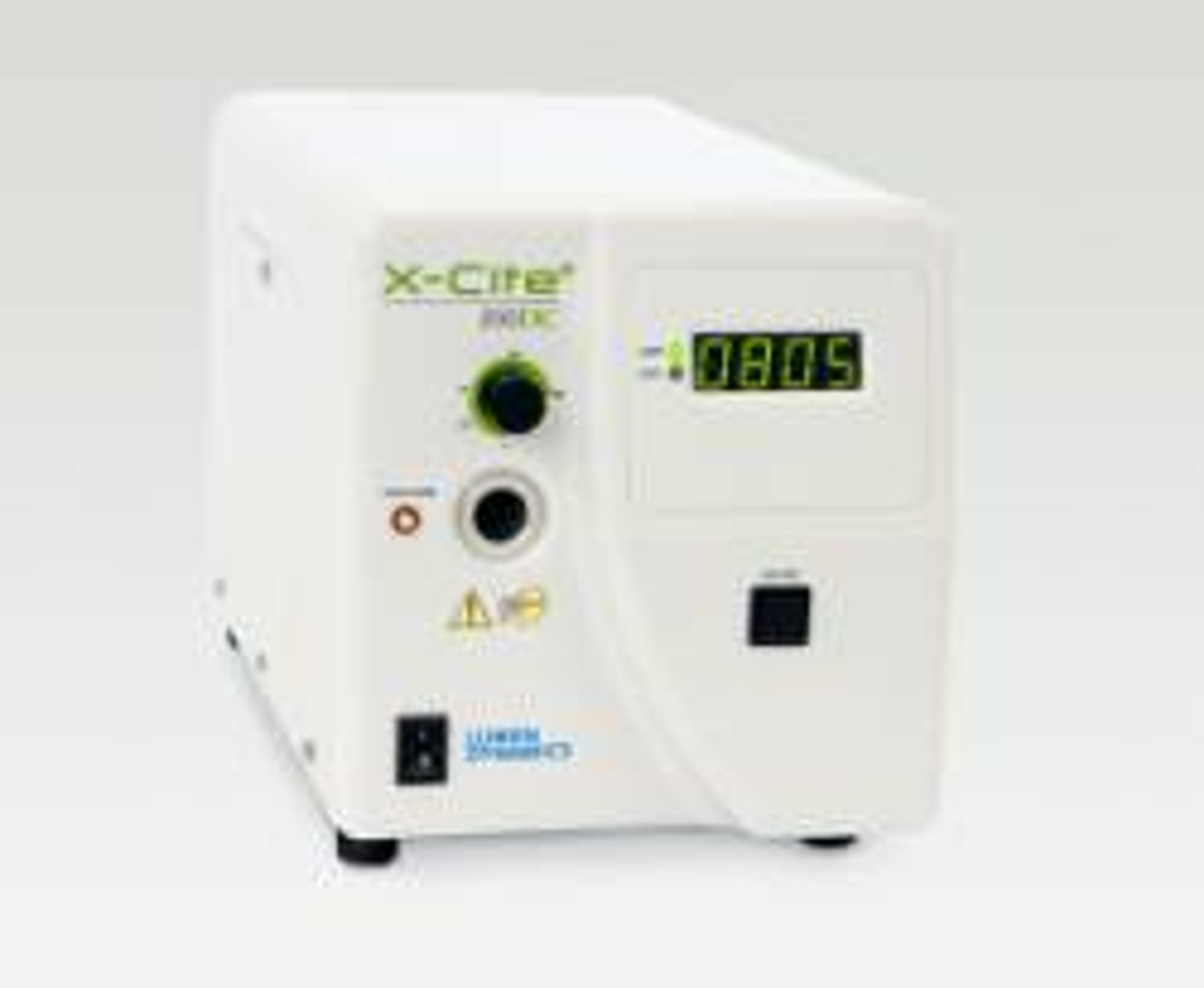
X-Cite® 200DC
Lumen DynamicsThe X-Cite® 200DC offers the ultimate combination of optical performance, short term stability and a built-in fast shutter. With the convenience and superior illumination uniformity found in all X-Cite® systems, the X-Cite® 200DC's intuitively easy-to-use design also includes light guide auto-detection to ensure optimized light coupling - every time. Novice and experienced microscopists will enjoy the flexibility provided by the smooth 0-100% intensity adjustment and expanded spectral range.