Looking for a compact microscope that works perfectly inside your CO2 incubator?
Introducing Curiosis’ automated live-cell imaging systems: The Celloger Series
8 Apr 2021
Product news

Beginning with the early discoveries of microscopes in the 16th century, there has been an endless desire to peer into objects that cannot be seen or are difficult to see with the naked eye. The development of technology and the introduction of live-cell imaging has transformed the way researchers study cells, tissues, proteins, and other cellular interactions, and has become a basic analytical tool in the study of life sciences. Live-cell imaging is a method to examine living cells over a period of time using images acquired by time-lapse microscopy. Through this technique, dynamic cellular processes and events can be observed in real-time during experiments to study and understand the biological changes occurring within cells.
Capturing live-cell images manually is a challenging and tedious task requiring researchers to exert much effort and time to acquire satisfying results. Difficulties in maintaining a suitable environment for the live cells to grow as well as keeping precise time intervals and sample positions during image capture makes live-cell imaging tough work. To bring an end to these complexities, Curiosis has launched a new range of user-friendly automated live-cell imaging systems, the Celloger Series.
Each system suits different needs. Celloger Nano is the most compact system among the Celloger series and easily fits and functions perfectly within a standard CO2 system. Moreover, it is equipped with exceptional fluorescence and auto-focusing technology, a precise stage controller, and intuitive software. Celloger Mini is an automated live-cell imaging system based on bright-field microscopy with fully motorized stages. The system is compatible with various cell vessel types that users can choose from based on their research protocol and multi-position imaging is possible up to 96 wells. Celloger Mini Plus is equipped with advanced fluorescence and bright-field technology that provides the highest quality images and time-lapse videos. Various cell-based research workflows and applications such as wound healing assay, cell proliferation, cytotoxicity assay, confluency, and growth curve can be examined using this all-around system.
Celloger Nano
- One-color fluorescence (Green or Red) and brightfield imaging technology
- Compact size that easily fits into standard CO2 incubators
- High-quality system yet cost-effective
- Equipped with precise stage controller
- One-click easy way to make a time-lapse video
- Compatible with various cell culture vessel types
- Intuitive UI/UX and easy to acquire confluency data
- Increased focus speed with reliable autofocusing function
Celloger Mini
- Live-cell monitoring and time-lapse imaging based on bright-field microscopy
- Compact size that easily fits into standard CO2 incubators
- Fully motorized stages and multi-position imaging up to 96 well plates
- Compatible with various cell culture vessel types
- Intuitive UI/UX and easy to acquire confluency data
- Increased focus speed with reliable autofocusing function
- One-click easy way to make a time-lapse video
Celloger Mini Plus
- One-color fluorescence (Green or Red) and bright-field imaging technology
- Compact size that easily fits into standard CO2 incubators
- Fully motorized multi-position imaging up to 96 well plate
- Compatible with various cell culture vessel types
- Multiple focal planes can be captured through Z-stack imaging
- Intuitive UI/UX and easy to acquire confluency data
- Increased focus speed with reliable autofocusing function
- Image stitching feature to enable analysis of larger volume and sections
Do you use Curiosis products in your lab? Write a review today for your chance to win a $400 Amazon Gift Card>>
Related products
Request Quote for All Products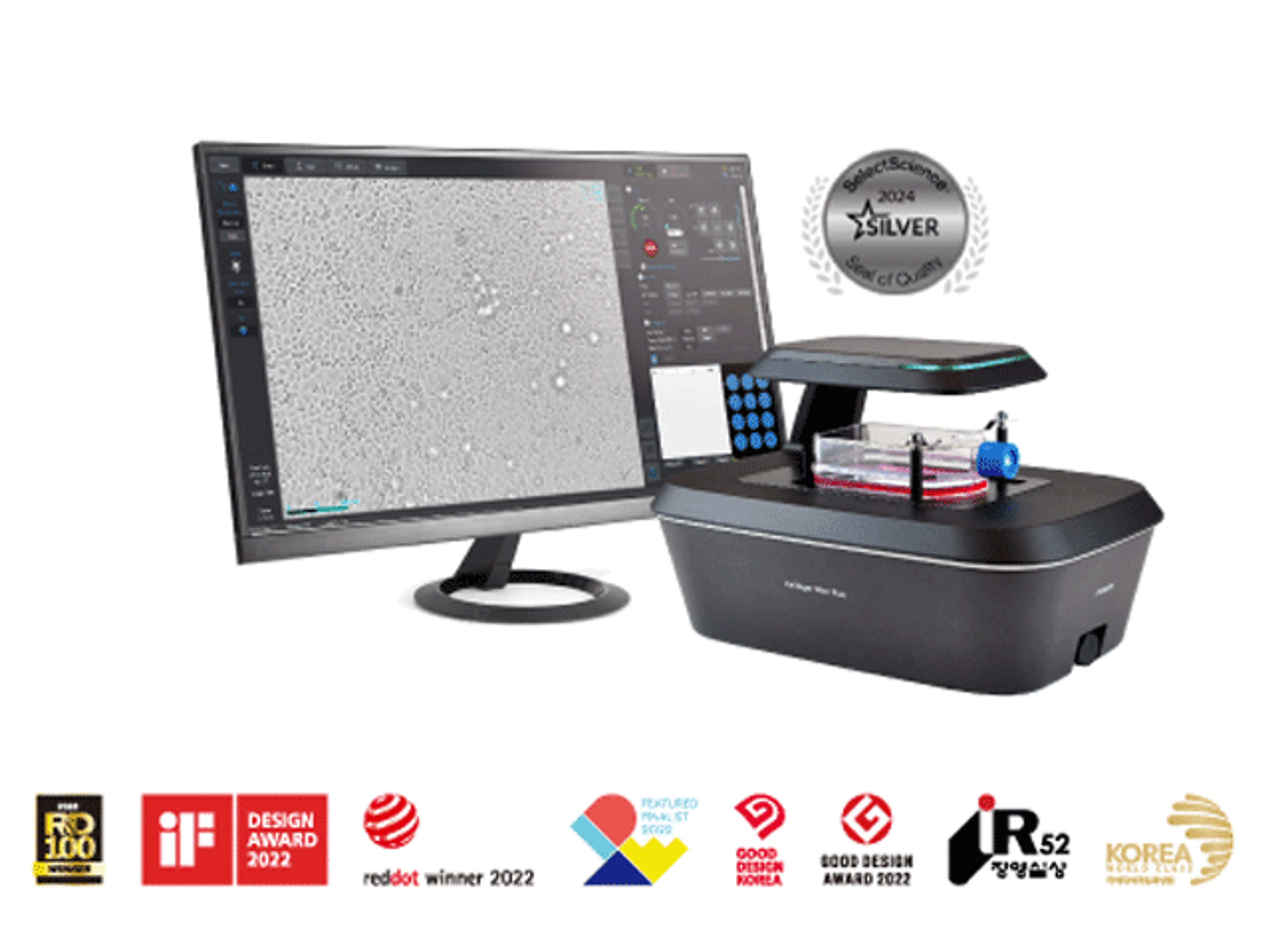
Celloger® Mini Plus, Automated Live Cell Imaging System from Curiosis
CURIOSISCelloger® Mini Plus is an automated live cell imaging system with fluorescence and brightfield microscopy. Celloger® Mini Plus makes it faster and easier to accumulate outstanding research results tailored to your research protocol.

Celloger® Nano, Benchtop Digital Microscope from Curiosis
CURIOSISCelloger® Nano is a benchtop digital microscope equipped with a wireless connection, enabling you to check the state of your cells in real-time from any location within your laboratory. With all the necessary functions to check the cells, you can quickly assess the condition of your cells.
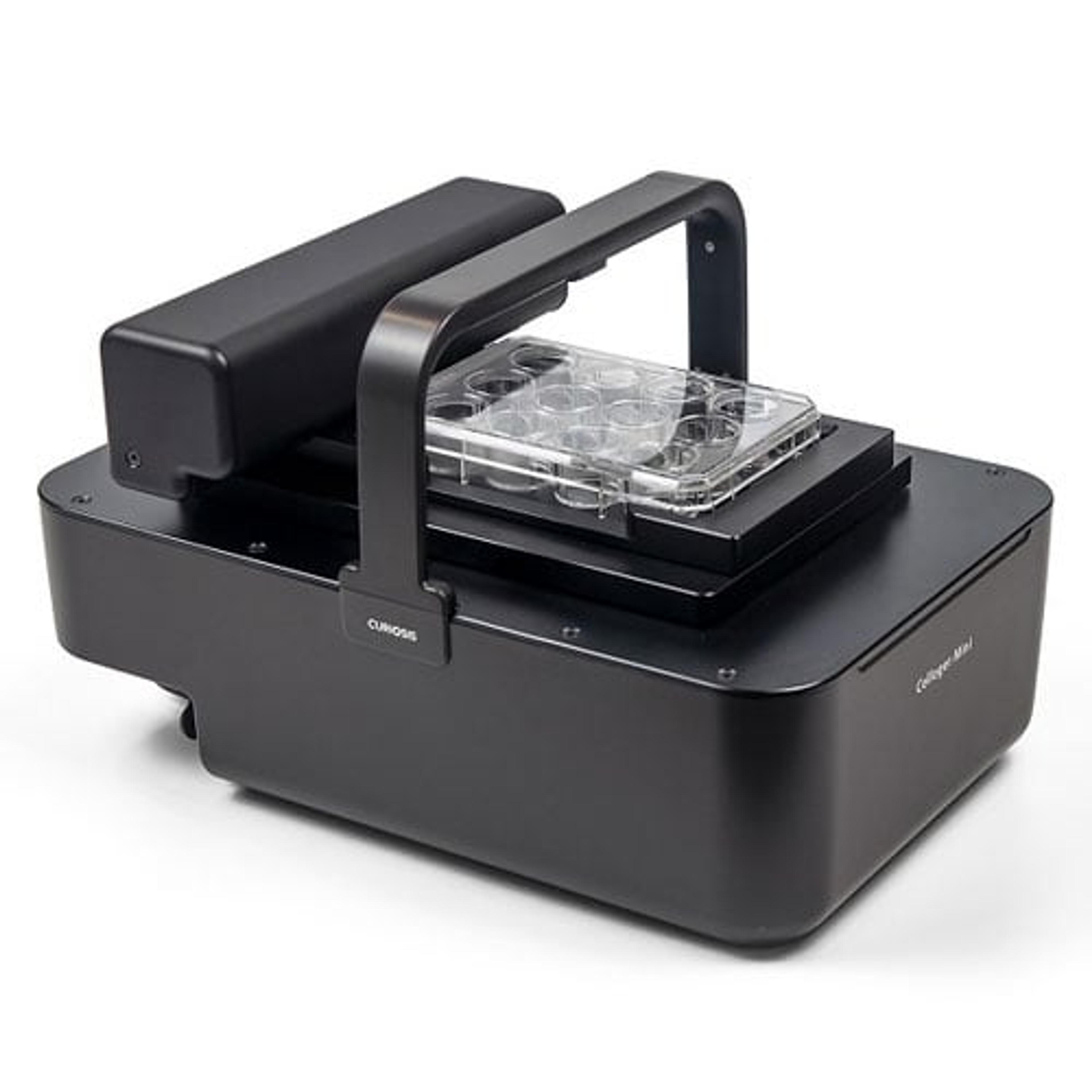
Celloger® Mini, Automated Live Cell Imaging System from Curiosis
CURIOSISCelloger® Mini is an automated live cell imaging system that uses brightfield optics with fully motorized stages to provide a precise, consistent, and user-friendly solution for the users.