Leica Microsystems launches ground-breaking Live-Cell CLEM workflow
The new solution promises to accelerate experimental success and improve process reliability as industry looks to faster drug discovery post-Covid-19
14 Jun 2021
Product news
Leica Microsystems has announced the launch of the Leica Nano workflow, a new live-cell correlative light and electron microscopy (CLEM) workflow solution designed to increase experimental success rates, improve reproducibility, and simplify light and electron microscopy (EM) integration.
The Leica Nano workflow combines high-contrast live cell imaging with EM sample preparation to provide a streamlined all-in-one workflow to answer a wide range of biological questions.
Understanding the basic building blocks of cells and tissue is a fundamental step in gaining better insights into the underlying mechanism and potential treatments of fighting diseases like cancer, neurodegenerative disorders and viral infections such as COVID-19. Here live-cell CLEM is providing a more complete picture in contrast to most workflows that are limited either by the sample range, the transfer time or the sample conditions during imaging and fixation – increasing complexity and risk of failure.
The Leica Nano workflow uses an intuitive, semi-automated three-step system to minimize the time between functional live-cell imaging and cryo fixation of the sample. A sample transfer between the light microscopy and the high-pressure freezer takes only a few seconds and results in a cryo fixed specimen.
The time-specific sample transfer and fixation enables the investigation of dynamic events and link these to the nanometre resolution of the EM without compromising the health and integrity of the specimen.
On the light microscopy side Leica’s Thunder Imager provides state-of-the-art computational clearing technology to clean up the typical haziness of the 3D image. The Leica Nano workflow also introduces a new immersion objective corrected for sapphire glass provide optimal imaging conditions. The resulting images capture cellular functional and ultrastructural information that can be easily distinguished and integrated at the single-cell level.
Markus Lusser, President of Leica Microsystems, said: “Accelerating the pace of drug discovery and development to bring vital therapies to patients faster is one of our industry’s number one priorities as we look to the world beyond the COVID-19 pandemic. Everyone from the biggest biopharma companies to new biotech start-ups are striving to speed up science and are looking to innovative new technologies to provide the solution.
“The Leica Nano workflow will play a central role in helping to achieve this goal by reducing the complexities of workflows in the laboratory and helping scientists to achieve a reliably high experimental success rate for live cell CLEM – the only commercial solution on the market to be able to do this,” added Lusser. “We are extremely proud of this new solution – one of a number we have planned for launch this year – and excited to see the reaction its already getting from our customers and the marketplace.”
Want the latest science news straight to your inbox? Become a SelectScience member for free today>>
Related products
Request Quote for All Products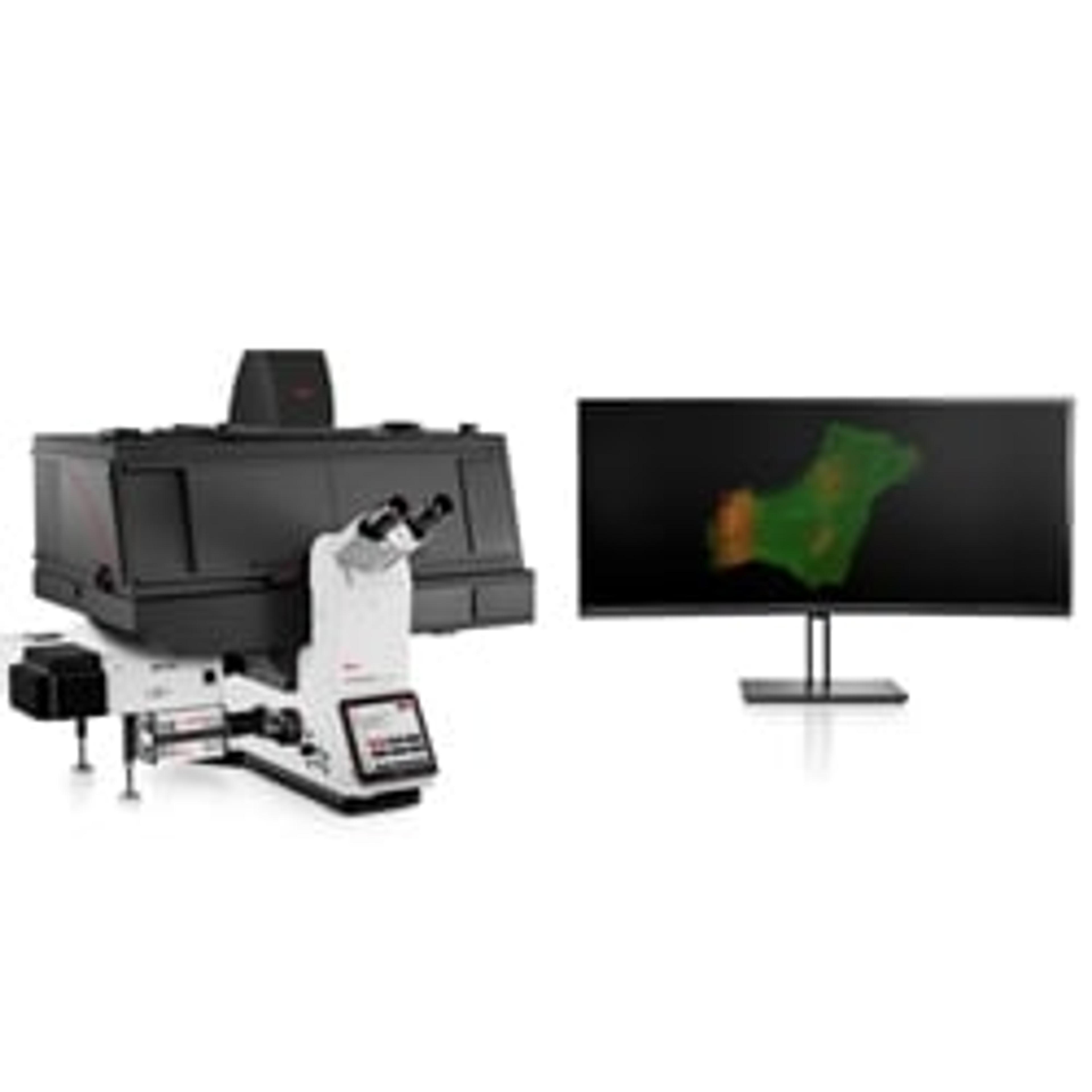
THUNDER Imager
Leica Microsystems EuropeYour solution for advanced 3D cell culture assays, whether you want to study stem cells, spheroids, or organoids