Launch of new Far-Red HI-LED lights from Syngene

9 Jun 2020
Georgina Wynne Hughes
Editorial Assistant

Product news

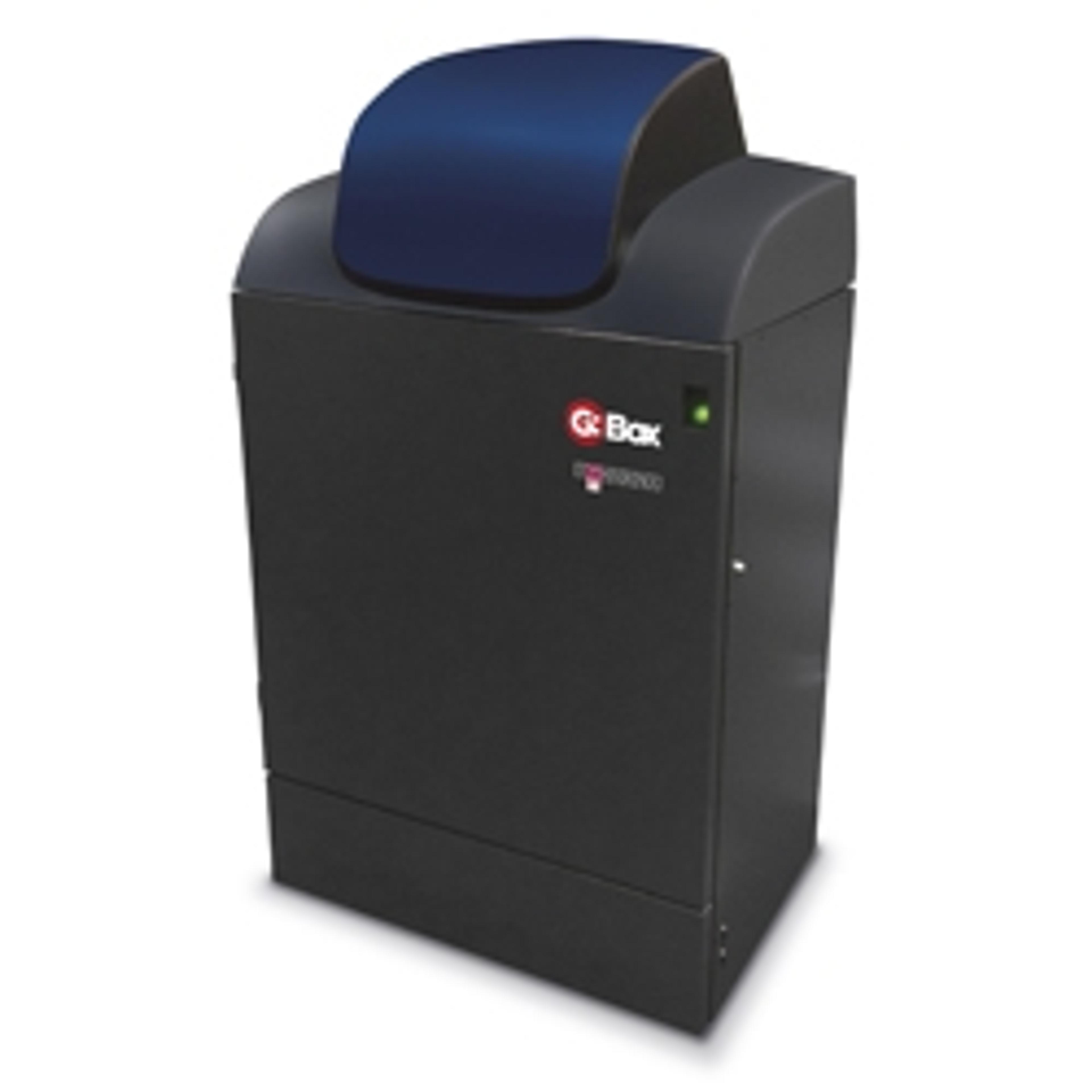
Syngene has introduced a far-red HI-LED lighting option for its new design of G:BOX Chemi and G:BOX mini multi-application gel and blot imaging systems. This quick-fit, environmentally friendly lighting enables fast workflow and precise detection of IR fluorescently labelled proteins on gels and blots.

With an excitation wavelength of 650-670nm, the far-red HI-LED light allows G:BOX Chemi and G:BOX mini systems to image sensitive near-IR protein dyes such as IR-Dye 680 and 700, and Alexa Fluor 680 and 700. The new LED light is also more intense than standard LEDs, which means that exposure times are shorter, making the G:BOX system into a high-speed imager.

The advantage of adding the new far red HI-LED light to G:BOX systems instead of purchasing dedicated IR detection technology, is that scientists still have the flexibility to image multiple different white and UV light applications. These include 1D/2D DNA and protein gels, as well as chemiluminescent and fluorescent multiplex Western blots and researchers can generate impressive publication-quality images using just one imaging system.

The eco-friendly, far red HI-LED lighting (up to 100,000 hours’ service life) is easy to fit and is automatically controlled via intuitive, icon-driven GeneSys software. This new near-IR lighting combined with the G:BOX system’s real-optical imaging power, provides unrivalled resolution and sensitivity comparable to most laser-based systems, allowing accurate detection (femtogram levels) of low-abundance proteins.

“Long exposure times using CCD imagers and not being able to justify buying laser-based technology means many scientists don’t use sensitive IR dyes, even though they would like to”, explains Dr Martin Biggs, Sales Manager at Syngene. “Our new far red HI-LED lighting is the ideal solution as it enables G:BOX Chemi or mini system users to work across a wider range of applications and detect low-abundance proteins without having to make major equipment purchases or learn new protocols.”

Want more of the latest science news straight to you inbox? Become a SelectScience member for free today>>

Links

Tags

Western BlottingWestern blotting equipment is used to transfer and identify specific proteins within a sample, reveal protein modifications, as well as give a semi-quantitative estimation of their concentration. Western blotting equipment includes all apparatus necessary to transfer proteins from gel to membrane and subsequent processing steps. Protein transfer can be performed by electroblotting with wet, semi-dry and dry transfer systems onto nitrocellulose and PVDF membranes. Blocking, washing and labeling of membranes follows, involving buffers, blocking reagents, blotting / incubation trays, labeling reagents, immunoblotting assays, antibodies and conjugates. Automated equipment for these steps is available to accelerate your lab workflow. Finally, detection and imaging of proteins can be conducted using gel documentation and imaging systems. Find the best western blotting equipment in our peer-reviewed product directory: compare products, check customer reviews and receive pricing direct from manufacturers.Gel Doc / Image AnalysisGel documentation (gel doc) or gel imaging systems are used for the analysis of proteins, antibodies and nucleic acid immobilized in polyacrylamide or agarose gels, membranes or microarrays. Explore a range of a gel imaging systems, densitometers, scanners, transilluminators or UV lamp + CCD cameras for your image analysis solutions. Colorimetric, fluorescent and/or radioisotopic samples can be visualized and documented for further analysis. See gel doc / Image analysis software for quantitative 1D and 2D analysis of your samples. Find the best gel doc / image analysis products in our peer-reviewed product directory: compare products, check customer reviews and receive pricing direct from manufacturers.FluorescenceThe emission of fluorescence occurs when a photon of energy is supplied to a fluorescent chemical compound by an external source, causing it to become excited. Fluorescence can be detected and measured for different purposes using microplate readers, fluorescence microscopes, fluorescence scanners, and flow cytometers.LEDImagingImaging techniques are essential for obtaining visual representations of samples to understand structures, processes, and function in biological, chemical, and physical research. These tools range from traditional light microscopy to advanced imaging modalities like MRI and electron microscopy, providing researchers with valuable data for diagnostics, drug discovery, and material analysis. Explore imaging solutions in our peer-reviewed product directory to compare products, check reviews, and get pricing directly from manufacturers.Chemiluminescence