Improve high-content screening images with award-winning objectives from Olympus Life Science
Capture higher quality multicolor images more quickly and achieve better quantitative results over the full field of view
16 Jul 2020Product news
The scanR high-content screening (HCS) station** version 3.2 delivers improved image quality, better quantitative data, support for challenging segmentation and classification applications and increased speed to help life science researchers maximize the information they get from their samples.
Better images for better data
Key to these functionalities is Olympus’ breakthrough lens manufacturing technology. X Line™ objectives offer simultaneously improved chromatic aberration correction, image flatness and resolution. This combination enables the scanR system to more rapidly acquire higher-quality multicolor images while achieving better quantitative results over the full field of view. Since the ability to get quantitative data from the scanR system is directly tied to image quality, X Line objectives help the system extract more information from images.
High signal-to-noise images
The v. 3.2 update brings support for Hamamatsu’s ORCA-Fusion sCMOS camera. The camera’s large 21.2 mm (0.83 in.) sensor chip captures more sample information with high quantum efficiency and sensitivity. This results in higher-quality images with minimal noise.
Expanded deep learning functionality
Using a self-learning microscopy approach, the scanR system’s AI functionality automatically analyzes data by incorporating a learned analysis protocol into its assay-based workflow. This improved functionality adds support for multi-class semantic segmentation, which enables it to tackle some of the most challenging segmentation and classification challenges in life science microscopy. Brightfield analysis, ultra-low signal quantification, label-free mitosis assays and the classification of complex phenotypes are all possible using scanR deep-learning technology.
These improvements in optics, camera support and software make the new scanR 3.2 highly suitable for high-throughput imaging and image analysis across a range of life science disciplines. It helps ensure that researchers can analyze large studies quickly, efficiently and without compromising on image or data quality.
**The scanR system is not sold in Japan.
Want the latest science news straight to your inbox? Become a SelectScience member for free today>>
Related products
Request Quote for All Products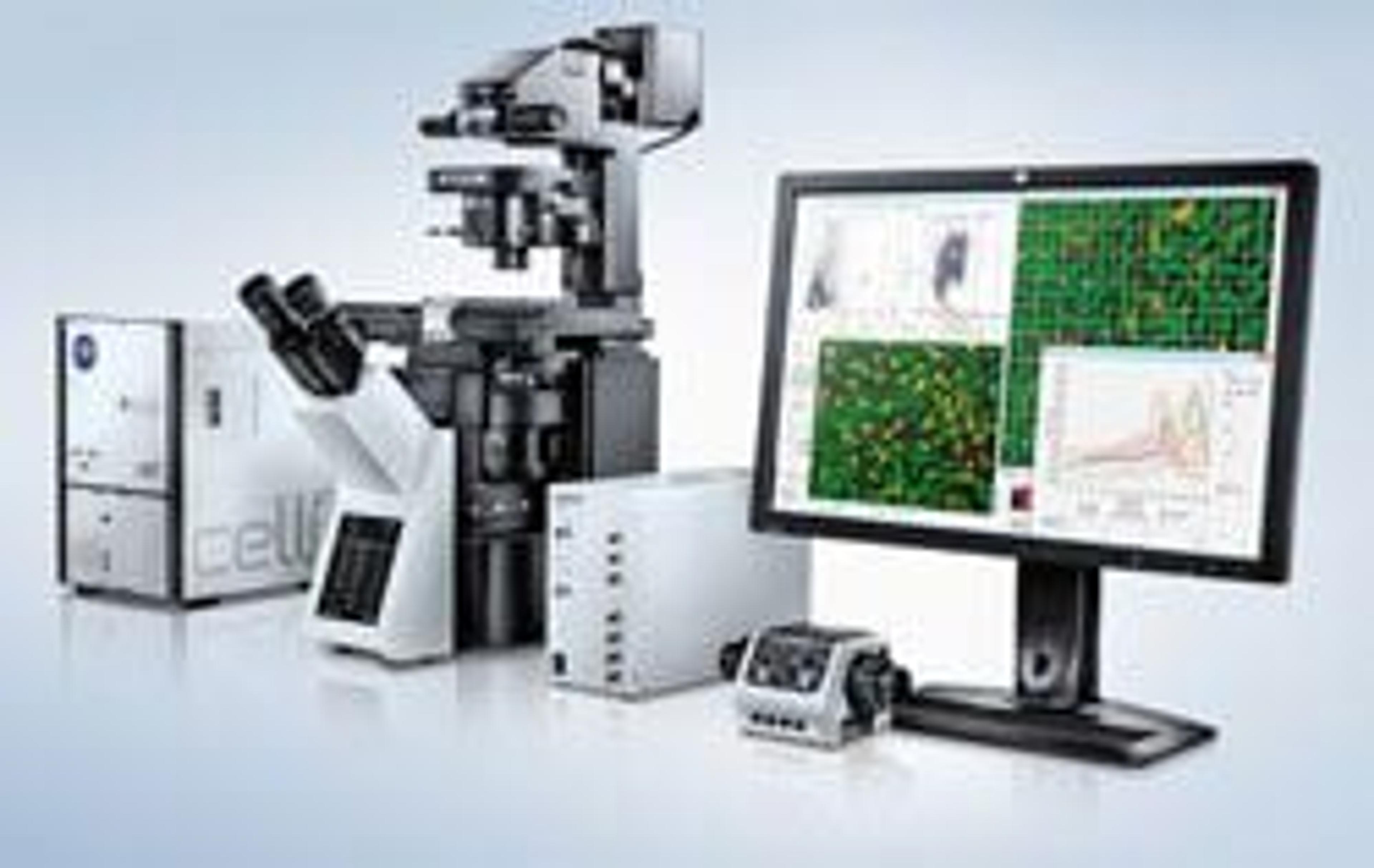
scanR - High-Content Screening Station
EVIDENTModular microscope-based high content screening solution