ibidi introduces the µ-Plate 24 Well for membrane inserts
Direct, high-resolution imaging of cells on membrane inserts without removal or disruption
8 Aug 2025Product news
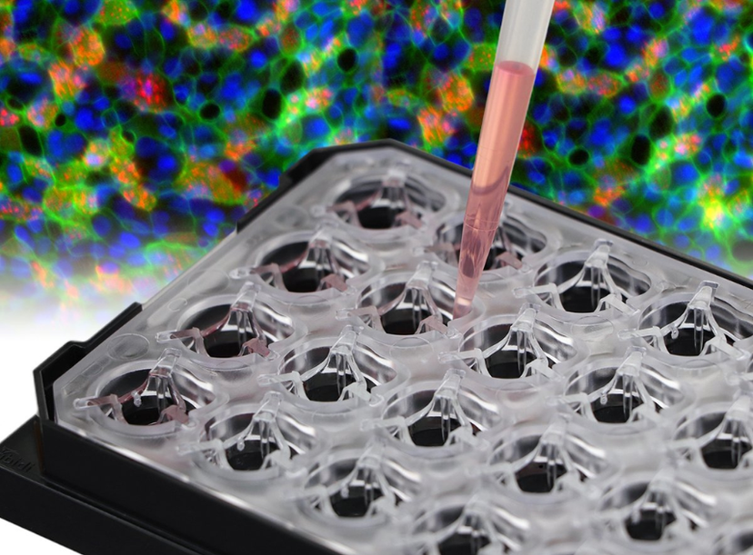
Cultivate, stain, and image cells grown on porous membranes—without removing the inserts. The new µ-Plate 24 Well for Membrane Inserts streamlines workflows and enhances imaging quality in cell culture experiments using standard membrane inserts.
Porous membrane-based inserts—also known as cell culture inserts, hanging inserts, or permeable supports—are essential tools in modern cell biology. However, traditional formats require the removal and manual cutting of membranes for imaging, which can cause sample disturbance, structural deformation, and imaging artifacts.
ibidi’s innovative µ-Plate overcomes these challenges with a two-position sliding mechanism, enabling seamless switching between cultivation, live cell and high-resolution imaging. During imaging, the insert can be positioned directly above the #1.5 ibidi Polymer Coverslip Bottom, which is optimized for microscopy. For continued cell culture, the insert can be easily returned to its standard hanging position—all without disrupting the cells.
The µ-Plate 24 Well for Membrane Inserts is compatible with standard 24-well-sized inserts from leading manufacturers including SARSTEDT, Greiner, SABEU, and Millipore and ensures easy integration into existing workflows.
Improve your membrane insert assay, including air-liquid interface cultures, barrier function assays, and co-culture models, while:
- Preserving sample integrity—direct imaging without insert removal prevents cell damage, reduces artifacts and allows for continuous cultivation.
- Enhancing reproducibility—standardized design and ease of use enable consistent results.
Do you want to test the ibidi µ-Plate 24 Well for Membrane Inserts in your experiments? Request your free sample at: ibidi.com.