How to use multiple observation methods to get the most from your slides

Industry expert in virtual slide scanning and upright microscopes shares an innovative combinatory approach to slide observation

5 Apr 2020
Diane Li
Assistant Editor

Expert insights

Dr. F. Ted Limpoco

Researchers often need to combine different observation methods to view structures that are only visible under certain conditions. In those instances where samples are in limited quantity, it is important to understand the combinatory approaches available in order to maximize the information that can be gained from any single slide.

In this on-demand SelectScience webinar, Alec De Grand, from Olympus Corporation of the Americas, discusses various observation methods to visualize a slide, including ways to combine methods in order to overcome one method’s disadvantage.

Read on for highlights from the live Q&A session or register to watch the webinar at any time that suits you.

Watch Webinar Now

Q: Do you need to combine methods to get the information?

ADG: The short answer is no, you don't always need to. Sometimes if you're just doing something like fluorescence where you just want to see your signal to see if they are a yes or no, that's enough. But if you want to add context to what you're looking at, maybe you want to add bright field or maybe you want to add dark field to the image to give you more information out of it. If it's for your everyday things, you may not need it. But for things like presentations or publications, you may want to try to push that level a little higher by adding another observation level.

Q: Are there any additional methods that can combine to gain more information?

ADG: Certainly. I talked about the major ones, but there are others, such as differential interference contrast or relief contrast/ Hoffman modulation or dot contrast. These are all variations on phase and polarization that also give you advantages and disadvantages. So, you could always mix those and match those as well. With fluorescence, now you're going to get confocal and light shading. These kinds of different observations that have very specific things, they also have disadvantages to them. There are many different observation methods when it comes to microscopy — you can mix and match. I just picked the most common ones that people would use a lot of.

Q: Can you combine these methods in whole slide imaging as well?

ADG: Yes. A lot of people do that all the time, usually bright field and fluorescence. Our system, like the VS200, can do all the methods that we're showing in the publication as well. So, they can mix and match. Normally two, sometimes three can be overlaid with each other. But when you overlay more and more observations, you can be blurring your data and not actually clarifying it.

Q: Are there automated slide scanners coming up with the ability to combine multiple imaging modes

ADG: Yes. As I said, the images that we're seeing here were on our VS series scanners, which can be achieved by mixing and matching. With the VS200, many systems have a bright field and fluorescence option. Not too many have the bright field, fluorescence and dark field. All of them kind of move together, but it's something coming down the road. There are a few out there that can do these things. Normally, it's an add-on option to add an extra observation method. But for the VS, it's built-in, except for fluorescence. That's an add-on.

Learn more about slide observation: Watch this webinar on demand >>

CX33 Biological Microscope

EVIDENT

Ergonomic and easy to use, our new CX33 microscope helps keep users comfortable during long periods of use, maximizing work efficiency.
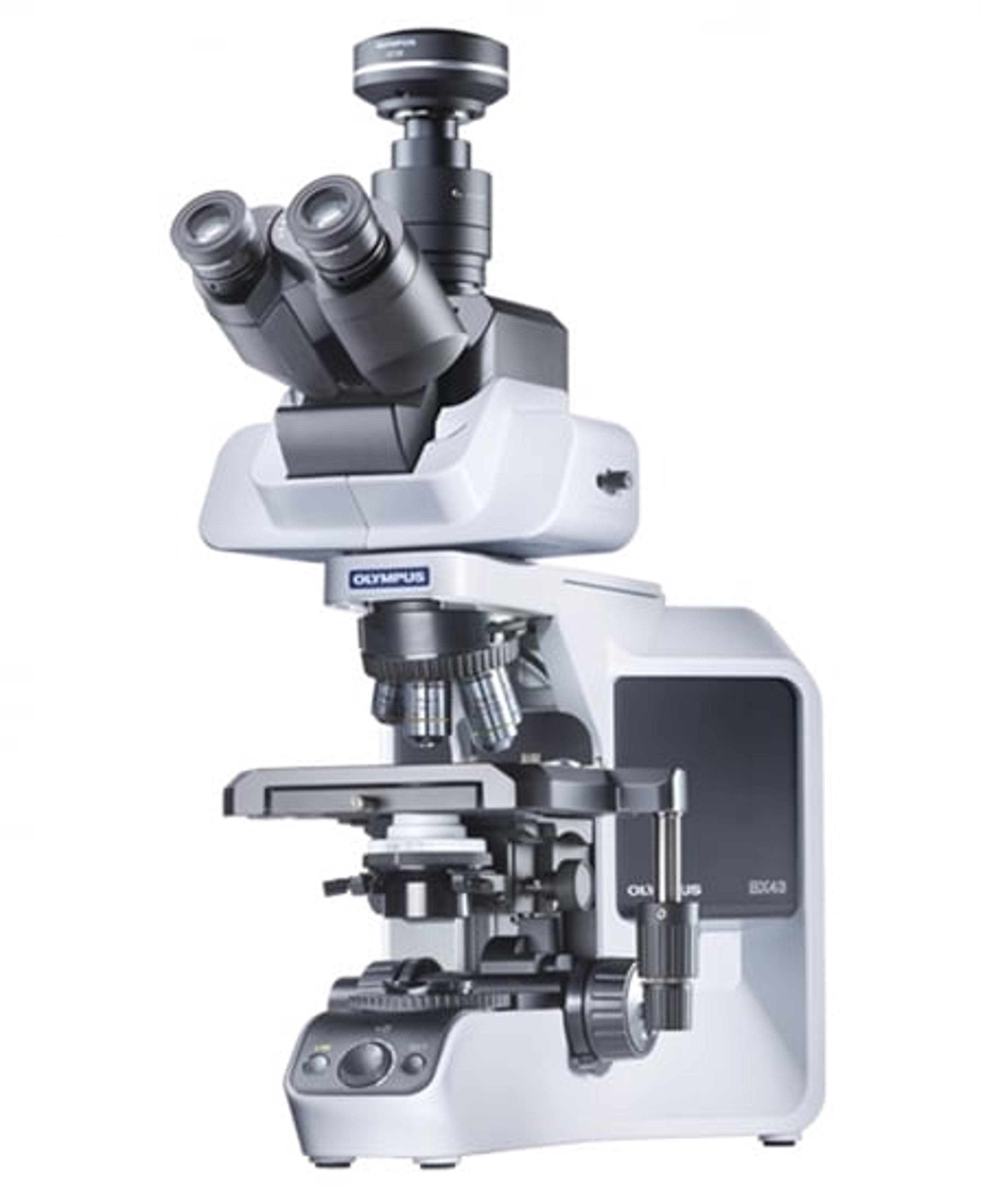
(1)

Links

Tags

In Vivo Imaging Systems<i>In vivo</i> imaging systems, including pre-clinical imaging systems and medical imaging systems are used to non-invasively visualize and capture images of live animals and plants. Monitor the natural processes or diseases of your subjects using small-animal pre-clinical imaging systems, including single photon positron emission tomography (SPECT), positron emission tomography (PET), computed tomography (micro-CT), magnetic resonance imaging (MRI), X-ray radiography, ultrasound, fluorescence and bioluminescence imagers. Multimodal systems and software solutions are also available for correlative analysis of organ, tissue, cell, or molecular-level processes. Find the best in vivo imaging products in our peer-reviewed product directory: compare products, check customer reviews and receive pricing direct from manufacturers.Light MicroscopyLight microscopes or optical microscopes are used to visualize microscale objects under magnification, including cells, clinical specimens and materials. Lab equipment for light microscopy includes confocal microscopes, fluorescence microscopes, zoom and stereo microscopes. Microscope slides and imaging reagents are available for visualizing samples, as well as various microscope stages and incubators for large or temperature-sensitive samples. Find the best light microscopes in our peer-reviewed product directory: compare products, check customer reviews and receive pricing direct from manufacturers.Electron MicroscopyElectron microscopes (EM) are used to create high-resolution images of samples at the nanoscale by means of an accelerated beam of electrons as a source of illumination. Types of electron microscope include scanning electron microscopes (SEM), transmission electron microscopes (TEM), scanning transmission electron microscopes (STEM) and cryo-electron microscopes. Focused ion beam (FIB) microscopes are useful for modifying or milling a sample surface with nanometer precision, as well as imaging. Find the best electron microscopes in our peer-reviewed product directory: compare products, check customer reviews and receive pricing direct from manufacturers.Digital MicroscopyDigital microscopy involves using digital cameras and sensors to capture high-resolution images of samples for analysis. It offers enhanced imaging capabilities compared to traditional optical microscopy and is widely used in biological and material science research. Explore digital microscopy systems in our peer-reviewed product directory; compare products, check reviews, and get pricing directly from manufacturers.Fluorescence MicroscopyFluorescence microscopy has become an essential tool in biology, as well as in materials science. The application of many fluorochromes has made it possible to identify cells and sub-microscopic cellular components with a high degree of specificity. Using multiple fluorescence labels, different probes can simultaneously identify several target molecules.ImagingImaging techniques are essential for obtaining visual representations of samples to understand structures, processes, and function in biological, chemical, and physical research. These tools range from traditional light microscopy to advanced imaging modalities like MRI and electron microscopy, providing researchers with valuable data for diagnostics, drug discovery, and material analysis. Explore imaging solutions in our peer-reviewed product directory to compare products, check reviews, and get pricing directly from manufacturers.MicroscopyMicroscopy is a technique used to observe small objects in detail, from cells to materials, using light or electron microscopes. It enables researchers to examine structures with high resolution, aiding in fields such as biology, medicine, and materials science. With advanced microscopy techniques, scientists can gain insights into cellular processes, tissue structures, and material properties. Explore the best microscopy solutions in our peer-reviewed product directory, compare products, read customer reviews, and get pricing directly from manufacturers.