High-Speed FRAP Imaging for Fast In-Vivo Processes
31 Aug 2015Product news
Olympus has introduced a flexible and accurate cellFRAP imaging platform specifically for the acquisition, monitoring and analysis of dynamic in-vivo processes by photomanipulation techniques. The easy-to-use platform delivers excellent optical performance and µs-precise control.
Enabling the easy addition of photomanipulation techniques to imaging platforms, Olympus has launched a new cellFRAP deck system for the popular IX3 ‘open source concept’ IX83 and IX73 microscopes. Olympus cellFRAP is designed with researchers in mind, providing highly accurate and flexible live-cell photomanipulation with various evaluation options for data presentation needs.
Unlike conventional widefield-based FRAP systems, Olympus’ cellFRAP employs a diffraction-limited laser spot for high-intensity and precise positioning of the bleaching region across the full field of view (FOV). This ensures that photomanipulation reaches every point in the FOV without needing to move the sample. The system’s excellent spot quality allows full flexibility in position and shape of the bleaching region, and enables control of the bleaching area with extreme accuracy. Driven by the Olympus Real-time controller, the cellFRAP system achieves an unrivalled short switchover time of only 200 µs between bleach and post-bleach acquisition, ensuring detection of the most valuable initial sample response after stimulation.
A chromatically corrected beam path enables multi-laser stimulation without the need for re-alignment, providing the ability to use up to four different lasers during the same experiment. This is especially beneficial for sophisticated applications such as optogenetics.
Via incorporation into the Olympus IX3 open source frames, the cellFRAP deck can easily be adapted to an existing imaging platform with a full range of compatible accessories including the ZDC, incubation chambers, manual or motorised stages and manipulators. This allows photomanipulation experiments to be set-up in combination with other high-end systems, for example, spinning disk or TIRF.
cellFRAP is seamlessly integrated into the Olympus Life Science imaging software, cellSens. A straightforward auto-calibration wizard makes it easy to adjust the scanner to the camera. Using the cellFRAP solution in the cellSens Graphical Experiment Manager provides intuitive system set-up and experiment control. This ensures excellent flexibility in experiment design and perfect synchronisation for maximum speed performance. Integration of any automated device is possible and even complex acquisition workflows can be easily defined and reproduced. The cellFRAP solution also includes a qualitative FRAP analysis for data evaluation. In addition, the Kymograph resembles timeline visualisation for easy extraction and presentation of data.
Tailored to the needs of photomanipulation, Olympus’ cellFRAP presents an ideal solution for researchers as an easy-to-use system that provides excellent optical performance, accuracy and µs-precise control.
Related products
Request Quote for All Products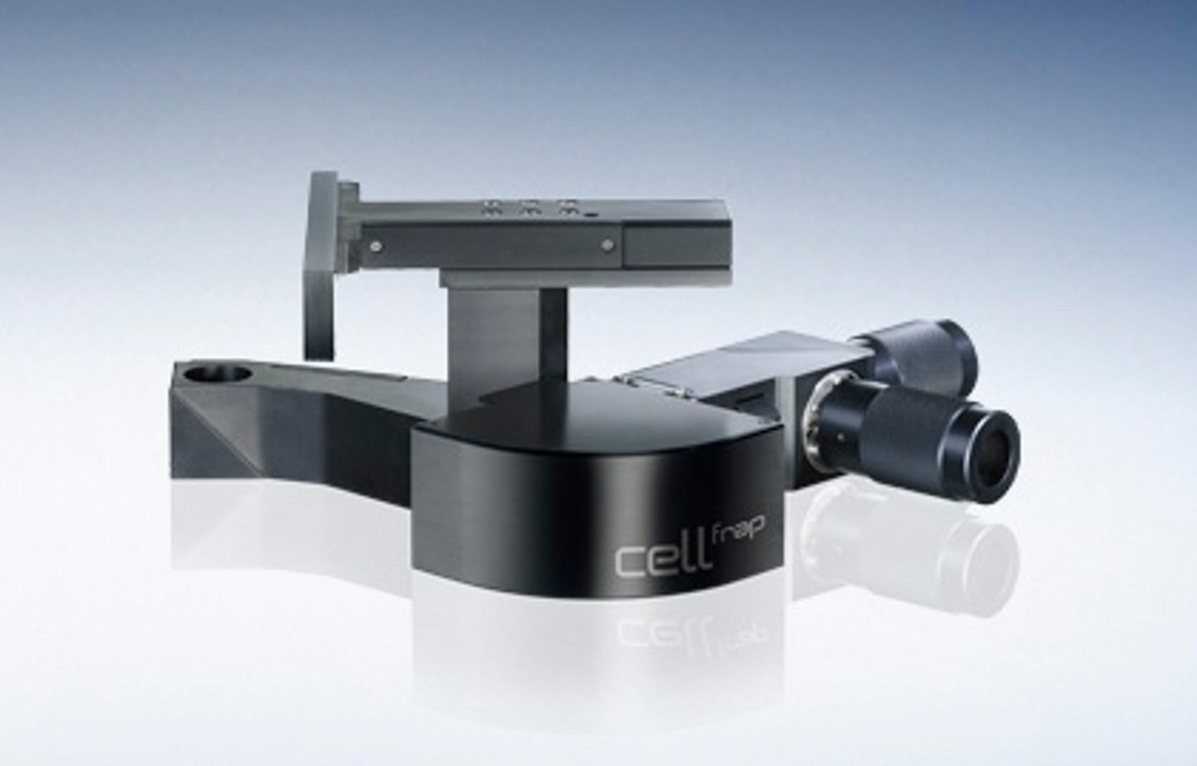
cellFRAP - Photomanipulation solution for Olympus imaging systems
EVIDENTThe cellFRAP system provides a versatile platform for a wide range of experimental needs, from basic bleaching to advanced protocols including FRAP, iFRAP, FLIP and FLAP. The system is also ideal for processes such as photo−conversion, photo−activation, pattern bleaching, laser cutting and trapping. An independent light path enables simultaneous imaging and bleaching and the module integrates seamlessly with other imaging modules and laser options. Intelligent scanning modes enable much faster bleaching rates than standard methods and facilitate accurate bleaching of very small structures. Features: From bleaching to photo−conversion - Users can control each setting for a broad range of procedures, including inverse FRAP, FLIP, FLAP, photo-activation, photo-conversion and photo-switching with multiple regions of interest. Optically advanced - The Olympus xcellence cellFRAP system uses an independent light path, which enables simultaneous fluorescence imaging and bleaching. Pattern bleaching - The cellFRAP system is capable of bleaching a large number of points in a defined pattern at high speed. Combined with diffraction limited optics, this method provides a useful and attractive alternative to fluorescent speckle microscopy (FSM). Rapid synchronisation - The Olympus cellFRAP module is capable of incredibly fast switching between bleaching and image capture, matching the requirements of all kinds of photomanipulation experiments. Flexible laser options - Can be integrated with two separate laser channels or a multi-laser combiner, providing excellent flexibility. Combined with UV-compatible optics, this gives the user an unmatched level of flexibility when it comes to laser and fluorophore choice. Intelligent scanning modes - By scanning only the region of interest, bleaching is achieved at a rate up to ten times faster than standard methods, and multiple areas within the same frame can be bleached very quickly. Sub−cellular laser cutting - The unique optical path employed by the system enables the use of pulsed lasers to cut cellular components such as growth cones and filopodia as well as intracellular structures such as actin fibres, microtubules, ER and mitochondria.