Gel and Blot Imaging Systems Now Offered with High Intensity Lighting for Stain-Free Imaging

9 May 2017
Lois Manton-O'Byrne, PhD
Executive Editor

Product news

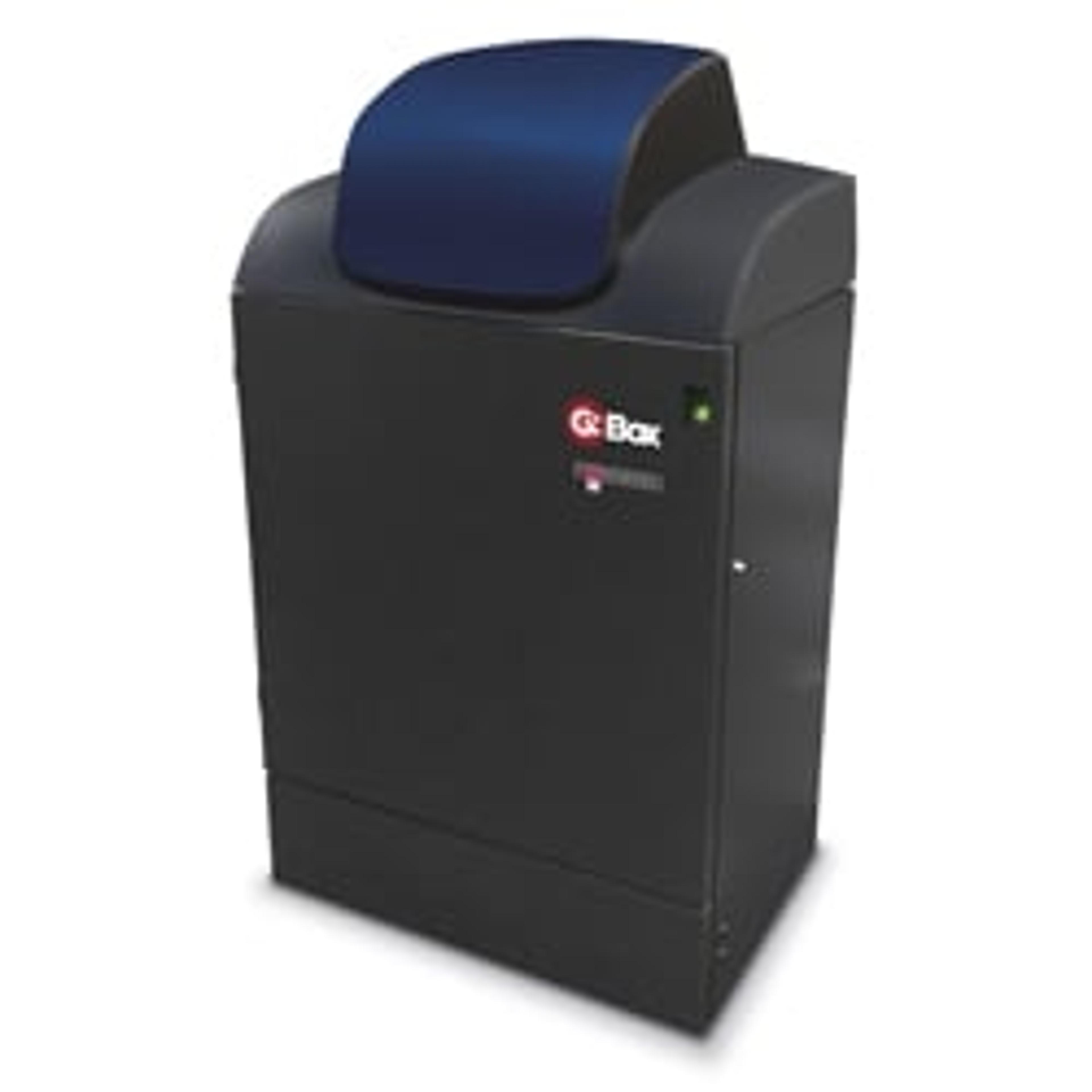
Syngene, a leading manufacturer of image analysis solutions, is delighted to announce its latest options for its highly successful G:BOX automated multi-purpose gel and blot imaging systems are now available. Utilizing high intensity, “HI-LED” lighting and updated image capture software, these flexible systems guarantee cost-effective imaging and faster workflow with a huge range of fluorescence gel and blot applications.

Featuring the option to add a full spectrum of high intensity blue, green, red and infra-red HI-LEDs that are up to 200 times brighter than standard LEDs, the new G:BOX options provide faster exposure times and great images in just one click. This makes the G:BOX systems an unrivalled, cost-effective alternative to expensive laser-based technology and offers a faster workflow than other CCD-based systems for imaging complex multiplex fluorescent gels and blots.

All systems in the G:BOX range are controlled via powerful new GeneSys software which now includes a simple icon selection of pre-set stain-free protein gel imaging conditions. The icon is based on optimum filter and lighting conditions that can accurately detect nanogram levels of protein on a stain-free gel and the software auto-calibrates to each gel or blot’s size to generate great publication-quality images every time.

The intuitive GeneSys software, which is fully integrated with the new G:BOX range, saves researchers time looking up recommended detection conditions by guiding them through set-up using a database of hundreds of commercially available dyes and stain-free options. This allows scientists to visualise gels and blots stained with, for example, ethidium bromide, Coomassie Blue and SYBR® stains, as well as imaging all types of stain-free gels, making accurate image capture incredibly easy.

“Many scientists want to image multiplex gels but don’t currently because it takes too long using a CCD-based imaging system or they cannot afford the laser-based technology to view them rapidly”, explains Dr Martin Biggs, Senior Divisional Manager at Syngene. “We’re excited to introduce our new G:BOX HI-LED options because these systems are capable of such incredible performance that they are perfect for any lab looking to access a quick, affordable method of rapidly generating high quality, chemi, fluorescence, colorimetric and now stain-free gel images time after time.”

Links

Tags

Western BlottingWestern blotting equipment is used to transfer and identify specific proteins within a sample, reveal protein modifications, as well as give a semi-quantitative estimation of their concentration. Western blotting equipment includes all apparatus necessary to transfer proteins from gel to membrane and subsequent processing steps. Protein transfer can be performed by electroblotting with wet, semi-dry and dry transfer systems onto nitrocellulose and PVDF membranes. Blocking, washing and labeling of membranes follows, involving buffers, blocking reagents, blotting / incubation trays, labeling reagents, immunoblotting assays, antibodies and conjugates. Automated equipment for these steps is available to accelerate your lab workflow. Finally, detection and imaging of proteins can be conducted using gel documentation and imaging systems. Find the best western blotting equipment in our peer-reviewed product directory: compare products, check customer reviews and receive pricing direct from manufacturers.Gel Doc / Image AnalysisGel documentation (gel doc) or gel imaging systems are used for the analysis of proteins, antibodies and nucleic acid immobilized in polyacrylamide or agarose gels, membranes or microarrays. Explore a range of a gel imaging systems, densitometers, scanners, transilluminators or UV lamp + CCD cameras for your image analysis solutions. Colorimetric, fluorescent and/or radioisotopic samples can be visualized and documented for further analysis. See gel doc / Image analysis software for quantitative 1D and 2D analysis of your samples. Find the best gel doc / image analysis products in our peer-reviewed product directory: compare products, check customer reviews and receive pricing direct from manufacturers.Protein PurificationProtein purification is a vital step in drug discovery, therapeutics, biotech and life science research. The purification process typically involves subcellular or membrane protein extraction with cell lysis kits, separation of proteins from cell debris by filtration or spin columns, and the isolation of proteins of interest from other proteins and impurities with affinity purification (including fusion protein tags and antibody binding proteins A, G and L), immunoprecipitation or chromatographic methods, such as ion exchange, size exclusion and immobilized metal affinity chromatography. All purification methods come in multiple formats for your laboratory needs, including agarose or magnetic beads, resins, columns and filter plates. Find the best protein purification equipment in our peer-reviewed product directory: compare products, check customer reviews and receive pricing direct from manufacturers.Gel ImagingGel imaging captures high-resolution images of electrophoresis gels to analyze DNA, RNA, and proteins. Essential in molecular biology, this technique helps visualize results for accurate quantification and documentation. Discover top imaging systems and compare user-reviewed options in our directory.DNA Fragment SizingSoftware SolutionProtein SeparationLabel-Free AnalysisMultiplexingMultiplexing refers to the ability to measure multiple targets or analytes simultaneously in a single experiment. This technique is valuable for high-throughput screening, diagnostics, and complex assays, as it increases efficiency and data quality. Browse our peer-reviewed product directory to find the best multiplexing products, compare tools, check reviews, and get pricing directly from manufacturers.