Fast Filter Wheel for Fluorescence Microscopy
2 May 2016Product news
Prior Scientific announces an addition to their range of microscope filter wheels and shutters.
The new HF110A/32 holds ten 32mm diameter filters and fits to the excitation ports of a range of popular microscopes from manufacturers including Leica, Nikon, Olympus and Zeiss.
Filtering of excitation and emission light is vital in fluorescence microscopy. The HF110A/32 filter wheel from Prior Scientific offers a way to quickly and precisely change the wavelengths of light that your sample is exposed to. The filter wheel takes less than 95 milliseconds to move between adjacent positions on the filter wheel, enabling the end user to quickly alter the fluorophore excited – ideal for multi-fluorophore applications where rapid imaging is essential.
If desired, it is also possible to mount (using an extensive range of adapters) the HA110A/32 filter wheel on the emission port of most microscopes. Magnetic covers on both sides of the HA110A/32 filter wheel allow quick and easy filter changing.
Both filter wheels and shutters can be easily controlled using the Prior Scientific ProScan® 3 control system, which is compatible with a wide range of microscope manufacturers imaging software packages. The end user can therefore control all aspects of their microscope system from a single point, allowing precise coordination of illumination, stage movement, focus movement, shutter and filter wheel operation and image capturing.
Related products
Request Quote for All Products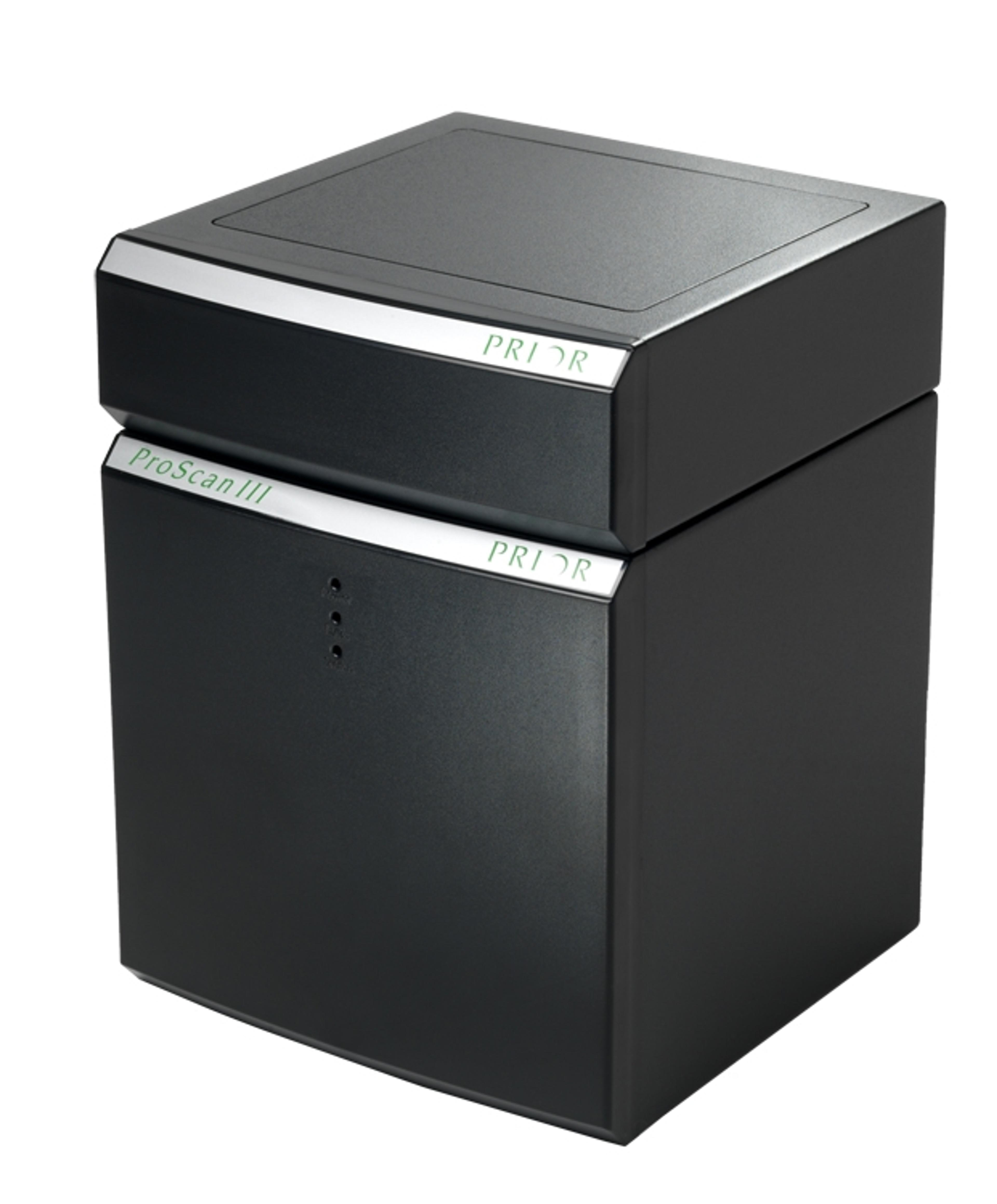

Filter Wheels
Prior ScientificThe filter wheels can fit to either the excitation or emission side of the imaging device. Magnetic covers over the filter load position on both sides of the filter wheel give easy access to allow filters to be changed quickly. Visible filter allocation numbers allow easy identification of filter positions, and a wide range of adapters for both illuminators and microscopes are available. In addition, Prior’s shutters can be mounted directly onto a filter wheel. The filter wheels are controlled via the ProScan® III control system. FEATURES: Easy to install and use 10 (for 25 mm filters), 8 or 6 (for 32 mm filters) position wheels available Compatible with almost all commonly used microscopes Controlled via Prior’s ProScan® III system. Adapters for a wide variety of microscopes and illumination devices available 55 ms change between adjacent positions on the filter wheel