Evolving High Content Screening with the Olympus IX83 Inverted Microscope Frame
20 Mar 2013
Product news
Olympus has expanded the scope of its most advanced IX83 inverted microscope frame, incorporating the scan^R 2.4 high content screening station for greater flexibility and increased scan speeds in a range of cell-based assays.
The scan^R 2.4 high content screening station is the latest addition to the growing collection of microscopy systems based upon the new IX3 high-end inverted microscope frame. Thanks to its unique “open source” design concept, the IX3 is offering a completely modular and flexible approach to live cell imaging. Built around a swappable deck design, optical modules can be easily exchanged into the accessible infinite light path, moulding the IX3 microscope to the diverse requirements of the user. The updated Olympus scan^R 2.4 high content screening station can now be incorporated with the most advanced model of the IX3 range, the fully automated IX83, combining benefits of a modular frame design with increased scan speed. This new system delivers quantitative expert results in a vast array of high content screening of cell-based assays, from gene expression to bacterial infection assays.
Keeping pace with the IX3 platform, the scan^R 2.4 software capabilities have also been extended, including fully supported multi-core analysis and unique kinetic analysis for advanced microscopy techniques. Furthermore, cell population data can be easily exported so users can jointly benefit from all the flexibility offered by complex experiments in the Olympus xcellence imaging software. The workflow-orientated graphical user interface has also been updated, allowing the advanced capabilities of the scan^R 2.4 station to be operated in a straightforward and intuitive manner, for routine and highly complex assays alike.
These new features build upon the performance of the scan^R stations’ high speed image acquisition and analysis. The dedicated real-time controller ensures precise timing of image capture, avoiding photobleaching and phototoxicity while maintaining the optimum scan speed. Designed to be flexible to user requirements, the scan^R 2.4 station is optimised for many different assay formats, including multi-well plates, slides and custom-built arrays.
Related products
Request Quote for All Products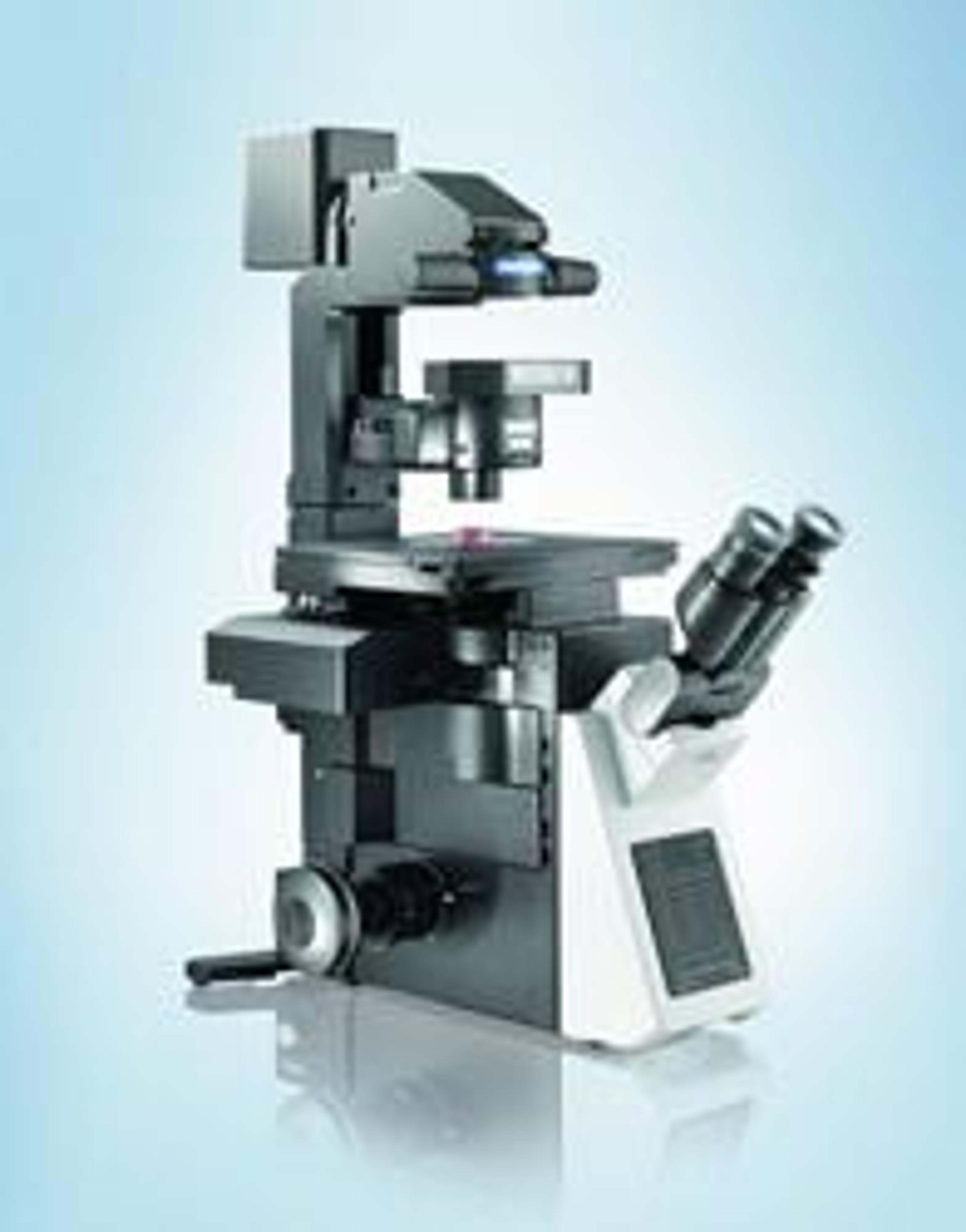
IX83 - Fully-motorized and automated Inverted Microscope System
EVIDENTExtended flexibility and performance in live cell imaging