Evolved Live Cell Confocal Imaging
7 Mar 2012Product news
Nikon Instruments has introduced the C2 plus, an enhanced version of its C2 Confocal Laser Point Scanning Microscope for entry and mid-level confocal imaging. Benefitting from faster scanning speeds, the new modular C2 plus also utilizes the latest version 4.0 of NIS-Elements software to provide increased accuracy and sensitivity in image capture.
Offering all the advantages of the C2, with its excellent hardware and software stability coupled with first class optics, the C2 plus provides improved galvano mode scanning speeds which are more than two times faster; up to a maximum 100fps (512x32) and 8 fps (512x512, bidirectional).
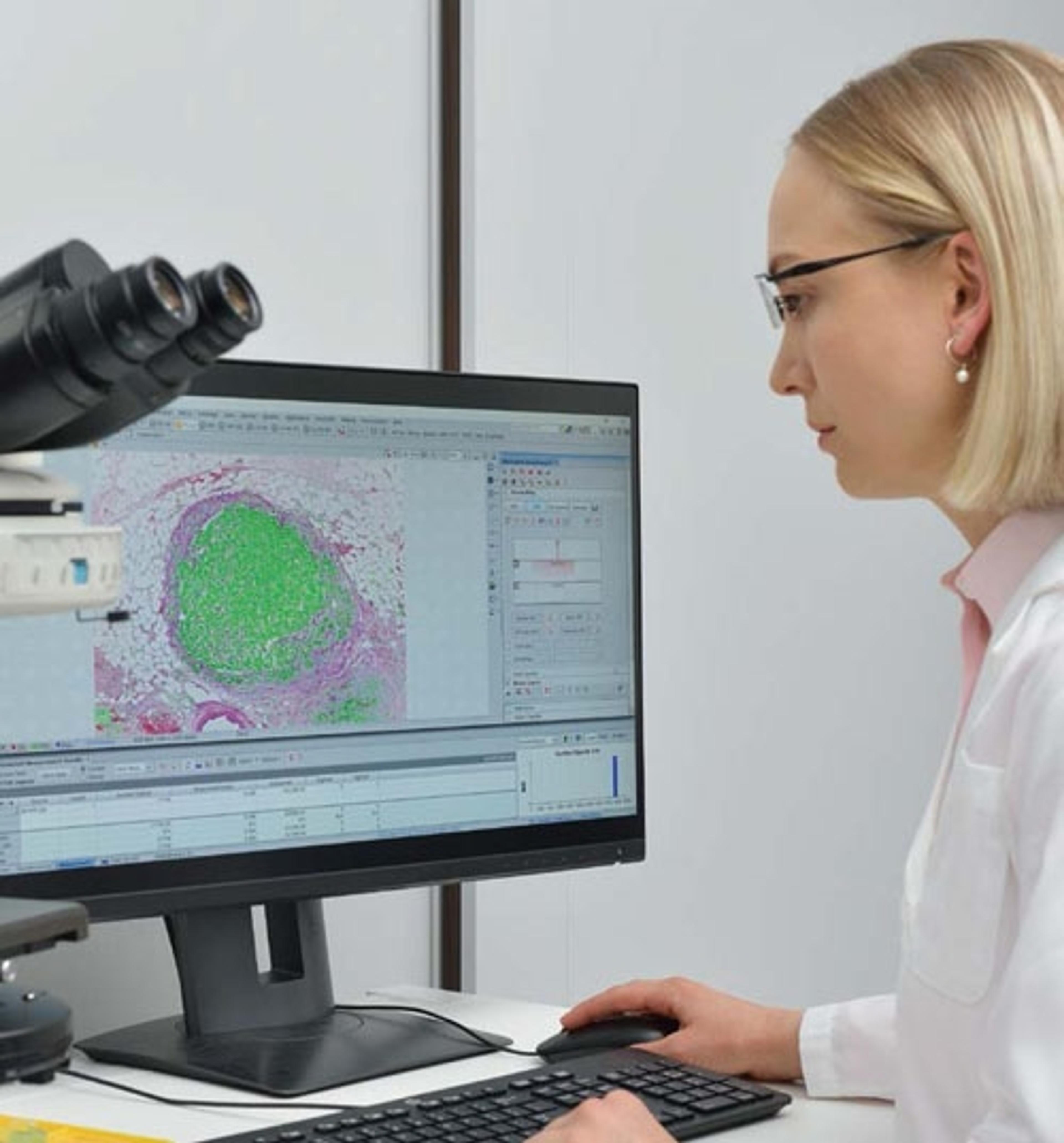
NIS-Elements version 4.0 now provides firmware control as well as improved image quality, dynamics and interaction, providing a consistent, universal interface and software environment for core imaging facilities and a variety of applications and techniques. Faster 3D image acquisition is possible, as well as improved high content screening functions for live cell observations and fully 2D interactive tracking.
The C2 plus can be coupled with Nikon’s new upright Eclipse Ni series as well as Nikon’s inverted, physiological and macro imaging microscopes, offering the advantage of just one universal software platform. Together with Nikon’s high numerical aperture CFI60 series objectives, optimized for live cell imaging with excellent resolution and brightness, they offer the ideal combination for confocal imaging.
Related products
Request Quote for All Products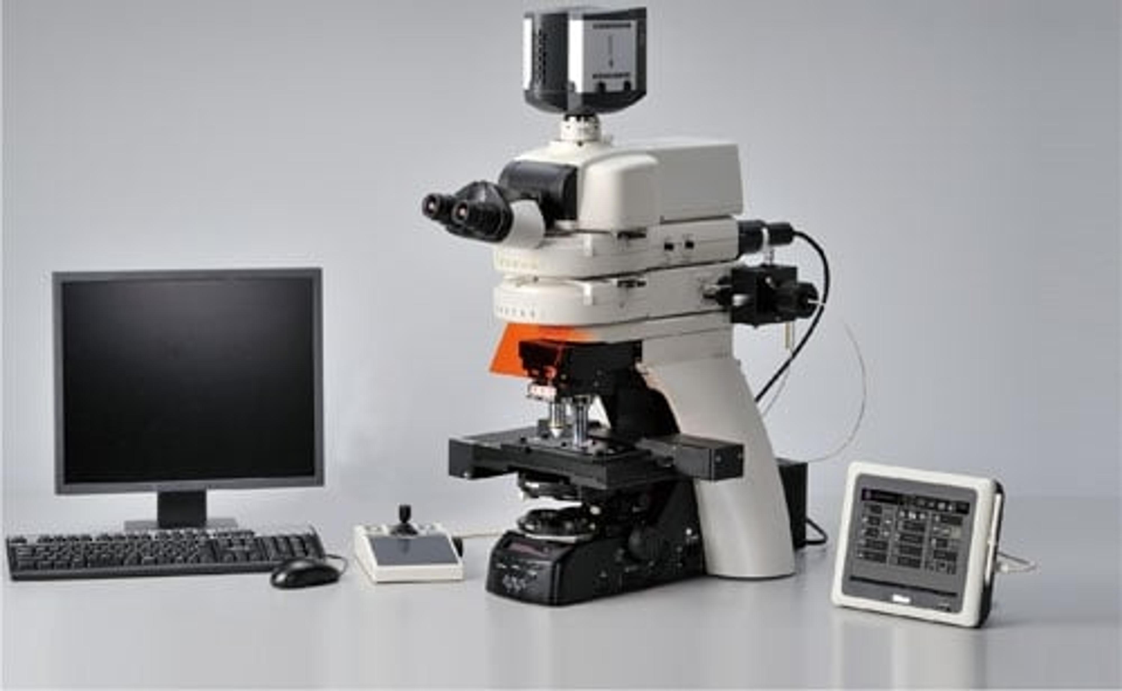
Eclipse Ni Upright Research Microscope
Nikon Healthcare Business – Microscope SolutionsResearch and clinical upright microscopes that support research in the fields of biological science and medicine, with excellent optical performance and high system expandability that can be equipped with motorized accessories.
NIS-Elements Microscopy Software
Nikon Healthcare Business – Microscope SolutionsFlexible software platform for controlling Nikon microscopes and 3rd-party components, with powerful custom programming tools for image acquisition and analysis.