DRAQ5 is an ideal DNA dye for fixed cell preparations in fluorescence microscopy
20 Nov 2007Product news
The far-red fluorescing nuclear counterstain DRAQ5, widely used for live cell imaging and flow cytometry, has also been validated by many recent publications for a wide range of fixatives, frozen and paraffin-embedded preparations.
Due to remarkable spectral properties DRAQ5 is compatible with the widest range of visible range fluorophores allowing simultaneous and multi-parametric imaging. One striking example is the combination of a constitutively-expressed GFP-tagged protein in female donor cells, a Y-chromosome FISH Cy3-probe for male recipient cells and DRAQ5 to demark the nuclei of all cells in mouse cardiac tissue sections (Visconti, 2006).
Usefully, DRAQ5 works with a full range of fixatives including methanol, acetone, PFA, and PLP and has been shown to stain efficiently both before (Schweitzer, 2005) and more typically after fixation (Grimm, 2004).
DRAQ5 penetrates deep into tissue sections: staining of 50 um-thick fixed sections from rat kidney samples (Toyokawa, 2006) permitted multiple confocal sections and 3-D reconstruction for a movie of three parameters.
DRAQ5 is compatible with wide-field, epifluorescent and confocal microscopes and all available HCS imaging platforms making it a perfect choice for broad application in immunohistochemistry.
Related products
Request Quote for All Products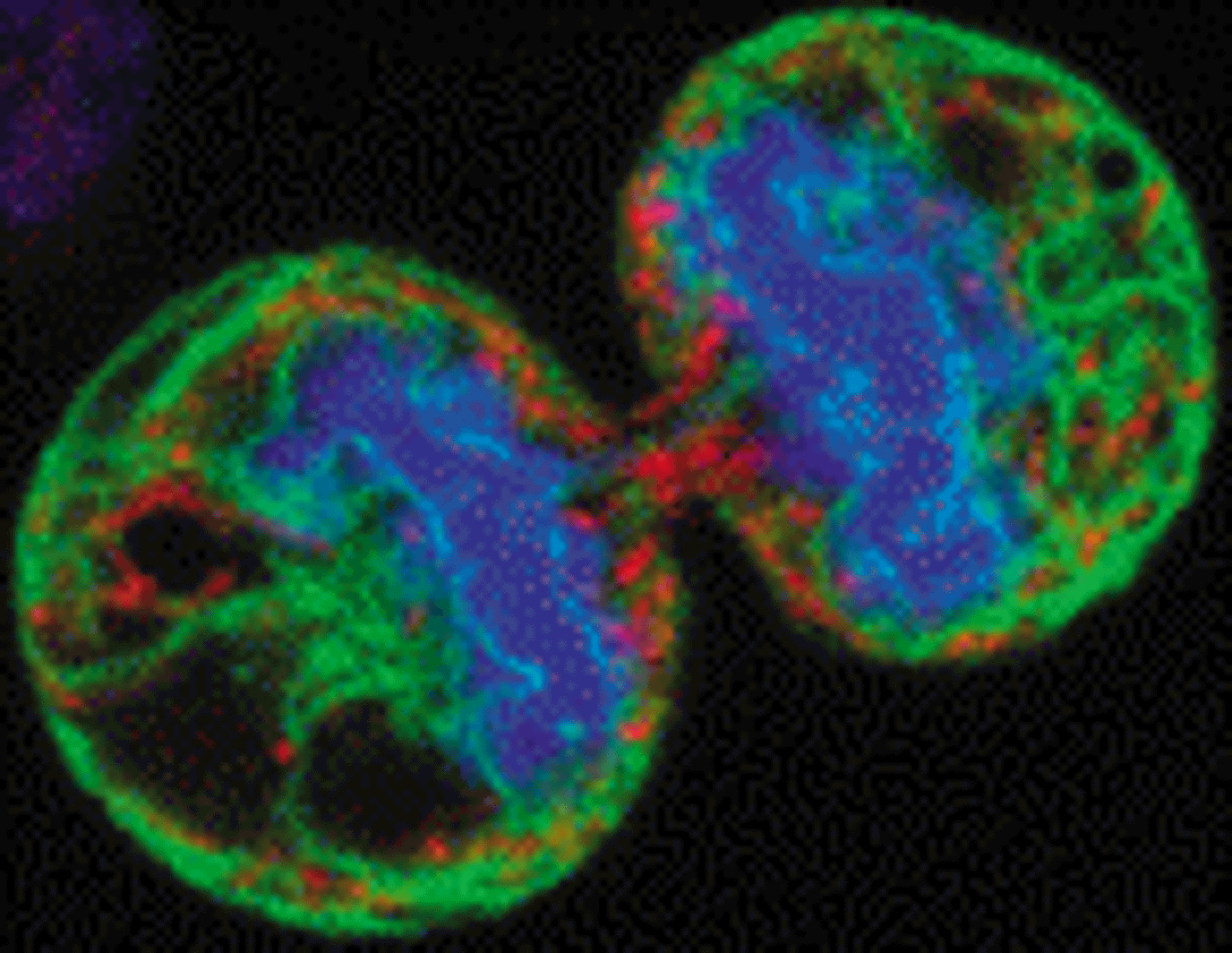
DRAQ5
Biostatus LimitedThe proven far-red probe for live or fixed cells, spectrally compatible with UV / Vis-range fluors.