CytoSMART Technologies announces the launch of its first fluorescence live-cell imager Lux3 FL
4 Nov 2020
Product news
CytoSMART Technologies has announced its first fluorescence live-cell imager. The CytoSMART Lux3 FL is a small live-cell imaging microscope equipped with one brightfield and two fluorescent channels (green and red).
The device enables researchers to unravel cellular processes in real-time, while the cells are kept in a controlled environment inside a standard cell culture incubator. CytoSMART’s first fluorescence live-cell imager allows users to track dynamic cellular processes with high specificity by taking high-quality images to create real-time time-lapse movies.
Said Jan-Willem van Bree, CTO at CytoSMART Technologies, "Currently, fluorescent labeling is mostly used as an end-point measurement. However, time-lapse imaging of live cells can give much more information about biological processes. By using automated imaging at regular time intervals, the temporal resolution of the fluorescent data is increased, leading to even more relevant data about the cellular processes. In this way, researchers can not only determine if a certain process has occurred, but also when it occurred and at what speed. Our customers have been asking us to develop a small and easy-to-use microscope with integrated image analysis of bright-field and fluorescence data. We have listened and made it happen."
The main features and benefits of the CytoSMART Lux3 FL include:
- Integrated image analysis of bright-field and fluorescence area or fluorescence object count.
- Time-lapse movies to investigate the development of cellular processes.
- Expanded number of variables researchers can analyze in their cell culture using green and red fluorescence.
- Remote data accessibility via the CytoSMART Cloud with a smartphone, tablet, or laptop outside the lab.
- Portable, easy-to-use and incubator friendly live-cell imager.
Do you use CytoSMART products in your lab? Write a review today for your chance to win a $400 Amazon Gift Card>>
Related products
Request Quote for All Products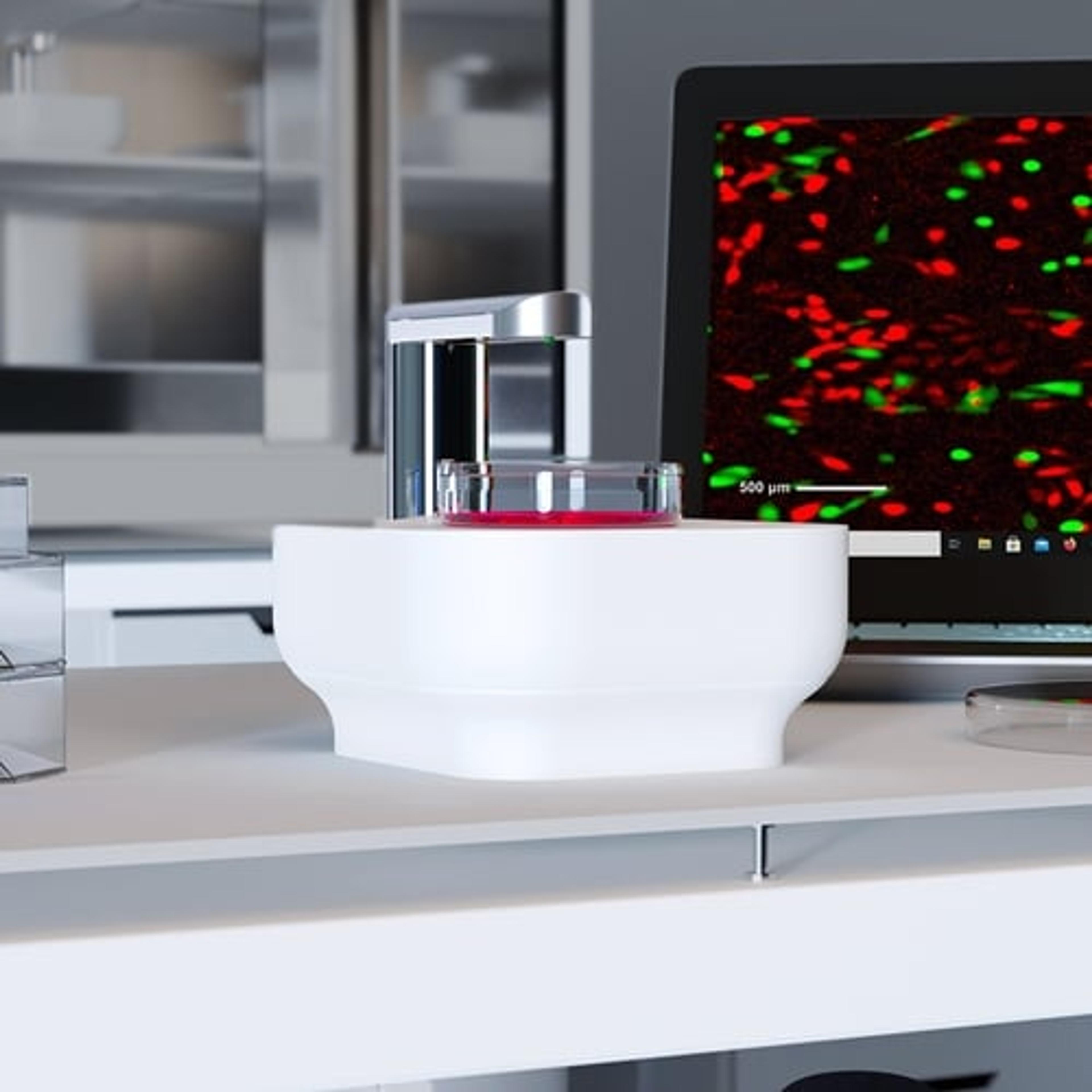
Lux
Axion BioSystemsSingle-well live-cell analysis with brightfield and fluorescent imaging from your incubator