CytoSMART introduces new, label-free live-cell microscopy solutions with high image quality
The CytoSMART Lux3 BR is a small brightfield microscope equipped with a high-quality 6.4 MP CMOS camera
9 Aug 2021Product news
CytoSMART Technologies has announced the launch of the new brightfield live-cell imaging system – the CytoSMART Lux3 BR. It is a small brightfield microscope, equipped with a high-quality 6.4 MP CMOS camera. The new live-cell imager is designed to work inside a standard cell culture incubator as all other CytoSMART microscopes, without disturbing temperature, airflow, and optimum culture conditions for the cells. This allows researchers to perform long-term live-cell imaging experiments, ensuring optimal cell growth and health. Very detailed brightfield images can be captured using the new device. In both x- and y-direction, 2072 pixels combined with 1.45 mm field of view provide a resolution of 0.7 µm/pixel. Even at the commonly required image resolution of 300 dpi for printed (scientific) publications, these images can fill the entire page width if desired, without compromising the image quality.
Said Jan-Willem van Bree, CTO at CytoSMART Technologies: “We are very excited to expand our label-free microscopy solutions for live-cell studies. For cell biologists who want to incorporate live-cell imaging into their workflow, the resolution of live-cell imaging microscopes may be too low for accurately quantifying complex read-outs, such as cell tracking or differentiation processes. This causes inaccurate and variable results that compromise the research output. Unlike the high-end microscopes with incubation box, the Lux3 BR ensures that cultures are not exposed to temperature, humidity, or CO₂ fluctuations that can stress the cells, as the device easily fits in every CO₂-incubator. It is a powerful addition to each research lab.”
A setup with the CytoSMART Lux3 BR can be easily expanded to two or even four devices that can be operated and controlled individually via a single laptop. Since both Lux3 BR Duo Kit and Multi Lux3 BR devices can be placed directly next to each other in the same incubator, it allows for systematic comparison of control and treated samples, as all monitored cell cultures are maintained in an identical culture environment.
The main features and benefits of the CytoSMART Lux3 BR include:
• High-quality images and time-lapse movies to investigate the development of cellular processes
• Incubator- and laboratory-friendly device due to its compact size and open design
• Remote data access via the CytoSMART Cloud with a smartphone, tablet, or laptop outside the lab
• Flexibility to expand a single live-cell imaging system to Duo and Multi Kits
For more of the latest science news, straight to your inbox, become a member of SelectScience for free today>>
Related products
Request Quote for All Products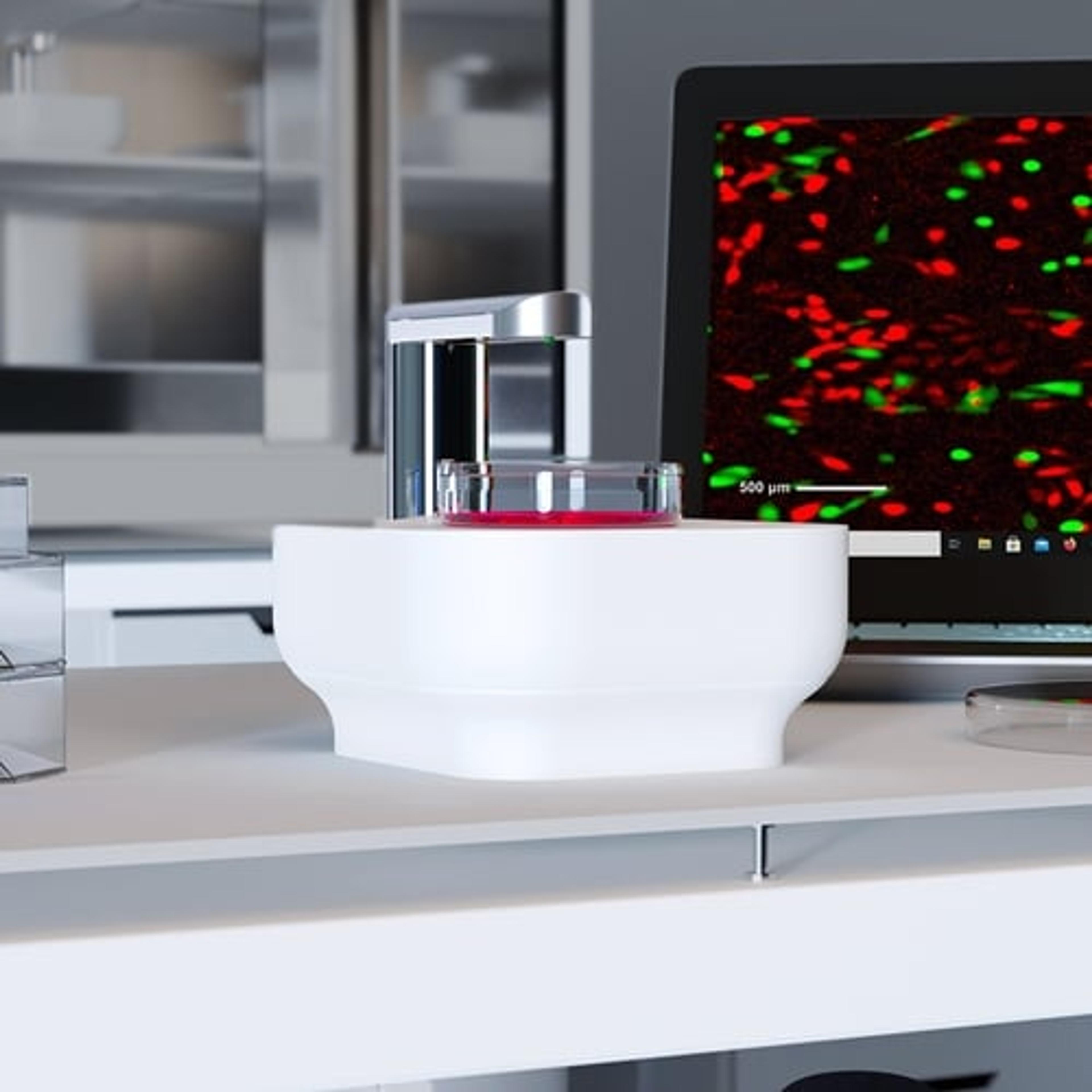
Lux
Axion BioSystemsSingle-well live-cell analysis with brightfield and fluorescent imaging from your incubator