CytoSMART expands long-term fluorescence live-cell imaging possibilities for simultaneous comparative studies
7 Jun 2021
Product news
CytoSMART Technologies has announced the launch of two new fluorescence live-cell imaging systems – the Lux3 FL Duo Kit and the Multi Lux3 FL. The CytoSMART Lux3 FL Duo Kit is a compact and cost-effective imaging system consisting of two fluorescence microscopes, designed for side-by-side comparison of cell cultures. The CytoSMART Multi Lux3 FL fluorescence live-cell imaging system consists of four compact Lux3 FL devices, equipped with two fluorescent (green and red) and one brightfield channel, and designed for long-term comparative studies and larger research teams.
Both systems operate from within a standard CO2-incubator or hypoxia chamber, allowing to analyze cells in their desired culture environment. Each system of two and four microscopes is connected to a single laptop, maximizing precious laboratory space. Monitoring and analysis of the running experiments can be done at any time from anywhere via the CytoSMART Cloud.
Said Jan-Willem van Bree, CTO at CytoSMART Technologies, "Last year we launched our first fluorescence live-cell imager, the CytoSMART Lux3 FL, and our customers were pleased with its performance and expanded research possibilities. This year we decided to scale up fluorescence live-cell imaging possibilities by launching two new advanced systems: the Lux3 FL Duo Kit and the Multi Lux3 FL. We always aim to develop and offer versatile and flexible solutions to life scientists. For example, the Multi Lux3 FL allows to run up to four individual imaging experiments simultaneously, without affecting the time-lapse imaging experiments of other lab members. This is a very convenient lab equipment for every research group that studies cell viability, co-culture models, single-cell migration, and many other cell-based assays can benefit from it."
The main features and benefits of the CytoSMART fluorescence live-cell imaging systems include:
- Ideal for comparative studies – directly compare fluorescently-labeled cell cultures.
- Versatile - expand the number of variables you can examine using fluorescent labeling.
- Flexible – connect devices to the same laptop and control them individually.
- Incubator-friendly – study cells in their desired culture environment.
- Full remote access via the CytoSMART Cloud - no need to enter the lab to inspect cell cultures.
- Indispensable tool for long-term comparative studies – run up to four experiments simultaneously for days or weeks.
- Cost-effective and efficient solution – two or four fluorescence imaging devices connected to one laptop, including unlimited storage and unlimited number of user accounts.
Do you use CytoSMART Technologies products in your lab? Write a review today for your chance to win a $400 Amazon Gift Card>>
Related products
Request Quote for All Products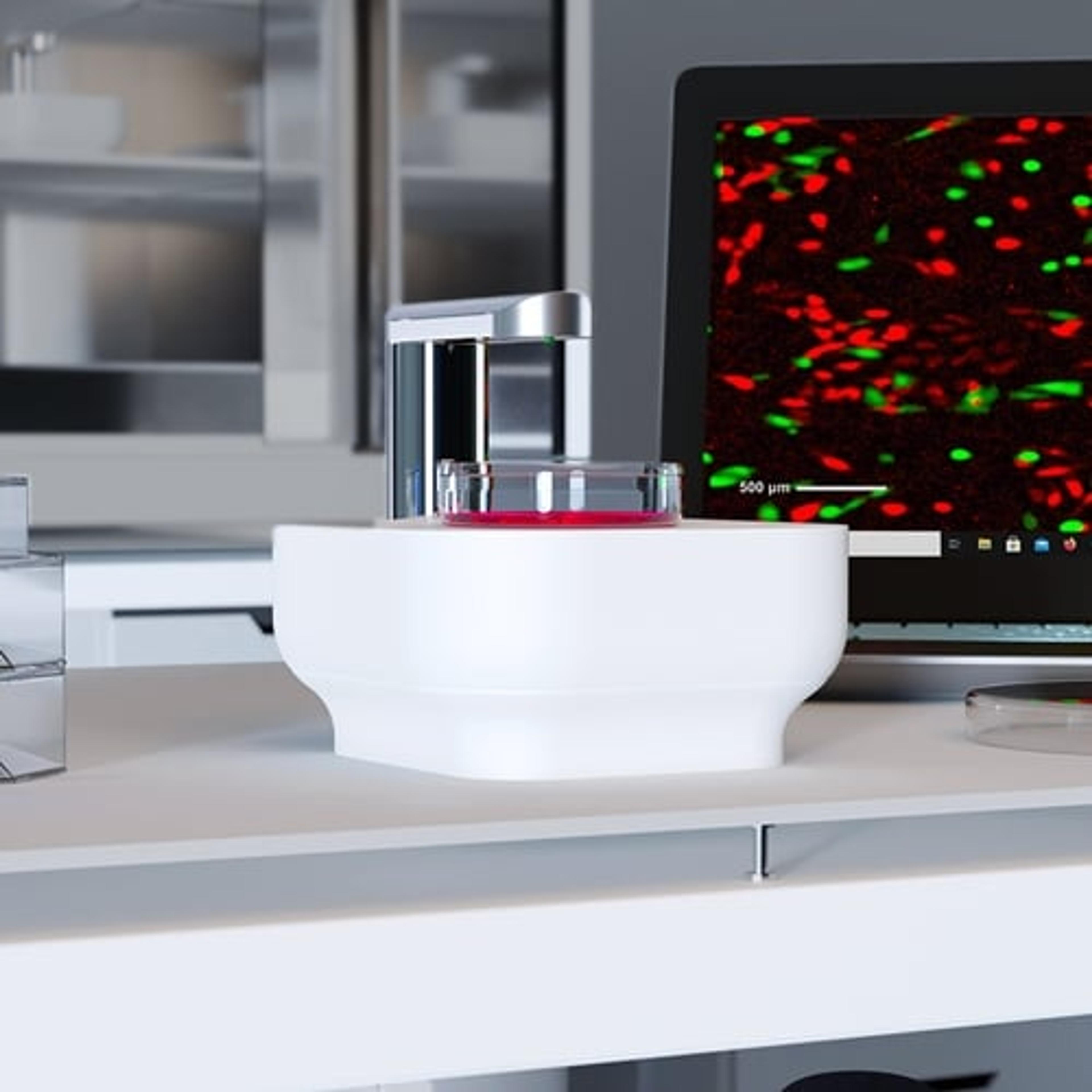
Lux
Axion BioSystemsSingle-well live-cell analysis with brightfield and fluorescent imaging from your incubator