CYTOO Cell Architects will be Introducing CYTOOplates™ at SBS 2010

5 Apr 2010
Sarah Sarah
Marketing / Sales

Product news

CYTOOplates™ feature thousands of adhesive micropatterns on an optical glass surface that reduce variability, simplify quantitative analysis, and improve reproducibility in HCA/HCS and other cell-based assays. Compatible with high content screening instruments, CYTOOplates fit seamlessly into your workflow. The poster entitled "Adhesive micropattern use in High Content Screening: a case study" will also be presented in location B-213.

To find out more visit booth 231.

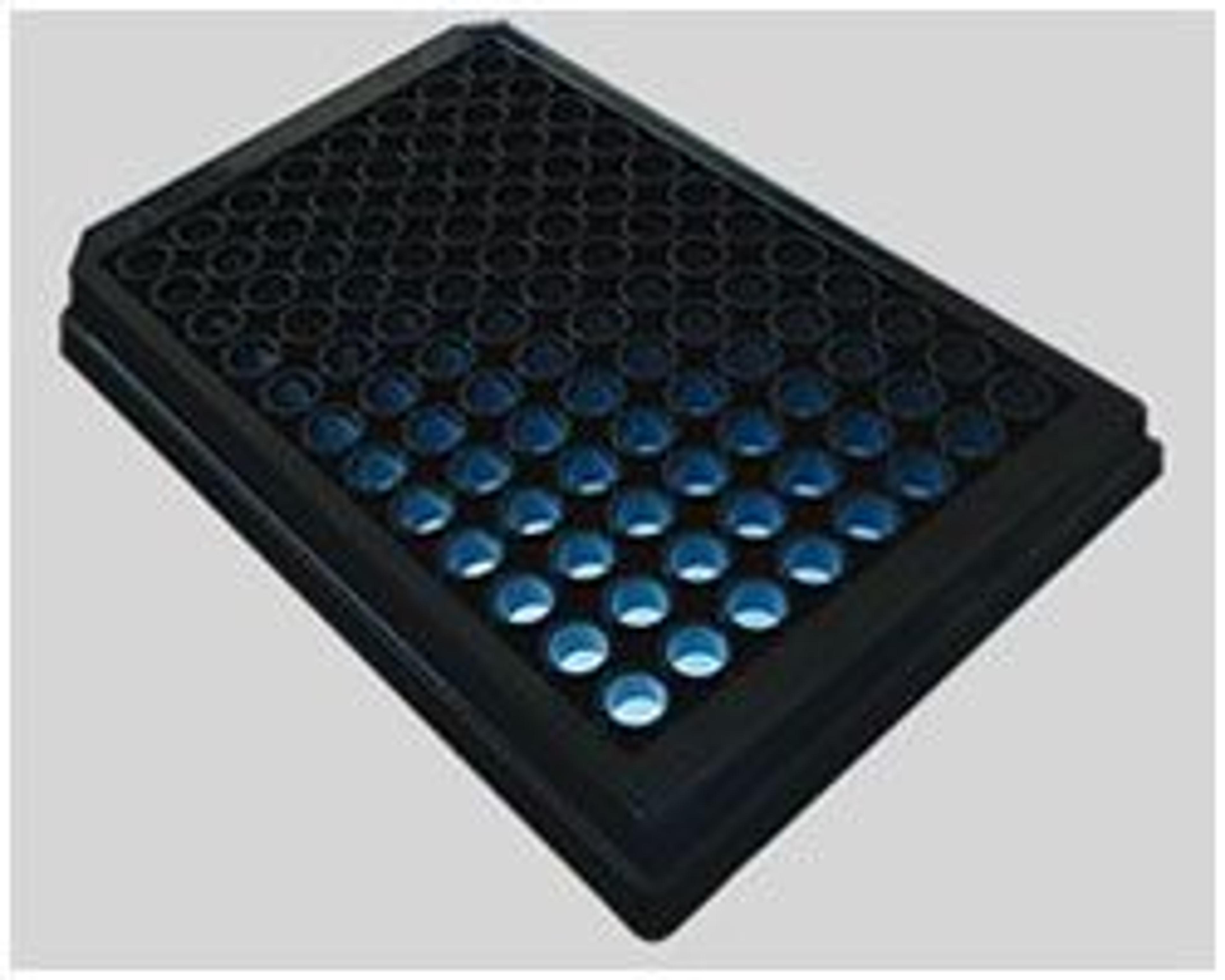
Control your Cells with CYTOOplates™

CYTOO Inc.

CYTOOplates are optical grade, glass-bottom microplates in 96 or 384-well format with black-side upper structures for fluorescence applications offering reduced background and minimum scattered light. Each well holds an array of over 4,000 (96-well) or 1,000 (384-well) identical micropatterns. CYTOOplates are compatible with your High Content Screening equipment.

(0)

Links

Tags

MicroplatesMicroplates are multi-well plates used to increase the throughput of biological investigations. The number of wells microplates contain can range from 6 to 3243 wells, with the 96-well format being the most commonly used. Microplates can come tailored for a range of applications including cell culture, PCR, filtration, storage, non-binding surface, protein crystallization, as well as pre-coated, deep well and normal Standard microplates. Additionally, options for microplate colors include clear , black, white or black with clear bottom and white with clear bottom for absorbance microplate reader applications. Find the best microplates for your lab in our peer-reviewed product directory: compare products, check customer reviews and receive pricing direct from manufacturers.High-Content ScreeningHigh-content screening (HCS), also known as high-content analysis (HCA), is a high-throughput technique used in drug discovery to identify substances that alter the phenotype of cells. HCS uses fluorescent microscopic imaging and automated image analysis to investigate cellular events such as apoptosis, cell viability, GPCR activation, oxide production, neurite outgrowth, and cell signaling. Find the best fluorescent labeling reagents, cellular assays, and high-content imaging systems in our peer-reviewed product directory: compare products, check customer reviews and receive pricing direct from manufacturers.