Carl Zeiss to Showcase Wide Range of Optical and Scanning Electron Microscopes at MICROSCIENCE 2010
21 Jun 2010Product news
Carl Zeiss will demonstrate the latest technology advances in SuperResolution, confocal and correlative microscopy, high-speed optical sectioning and digital imaging cameras and software. The ELYRA SuperResolution workstation will also be on display alongside the new VivaTome and ApoTome.
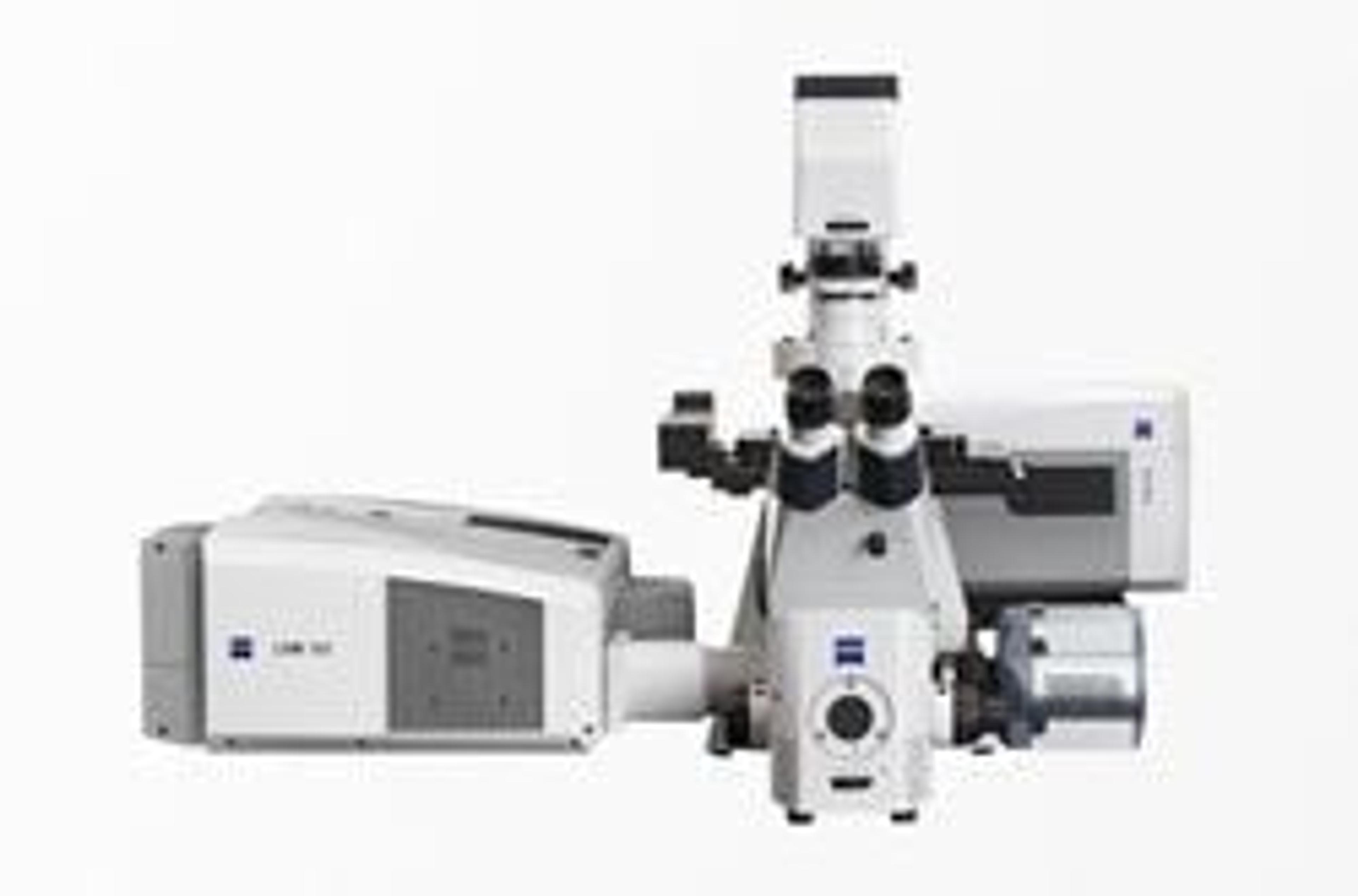
The ELYRA SuperResolution workstation will spearhead the display. Offering 20nm resolution, 10x greater than conventional light microscopes, ELYRA is set to revolutionise our understanding of the fundamentals of cell biology. Alongside the ELYRA workstation will be the groundbreaking LSM 780, which features radical, high quantum efficiency, GaAsP detector technology and offers double the sensitivity of the previously class-leading Zeiss LSM710. The LSM 780 will be demonstrated on the stand and the system also forms the platform of the new ELYRA SuperResolution microscopes.
Also on show will be the new VivaTome, pioneering optical sectioning technology that combines the imaging speed of a spinning disk system with the light efficiency of structured illumination. Designed for life science applications where temporal resolution is a priority, VivaTome provides developmental and cell biologists with a fast, cost-effective tool to examine the dynamics of living specimens without extensive prior knowledge of optical sectioning. Carl Zeiss pioneered optical sectioning through structured illumination with the award-winning ApoTome and this will also be on display.
Bridging the Micro and Nano worlds, Carl Zeiss will also be demonstrating its “Shuttle & Find” technology. This correlative interface for light (LM) and electron (EM) microscopes allows any point or region of interest (ROI) in LM to be relocated automatically at much higher resolution in the EM. Shuttle & Find is compatible with motorised versions of the Axio Imager, Axio Observer and SteREO Discovery LM platforms and all SEM/CrossBeam® electron microscopes.
Surrounding these revolutionary technologies, the Carl Zeiss stand will highlight the world’s largest array of light microscopes and digital imaging devices for the biomedical and material sciences. The all new Axio Lab.A1 microscope will make its UK debut, with versions that span the life science and materials worlds. Also on show will be the AxioCam ERc 5, a 5 megapixel camera that offers outstanding image resolution and colour accuracy, making it ideal for education and routine laboratory image capture.
Visit booth L8 to find out more.
Related products
Request Quote for All Products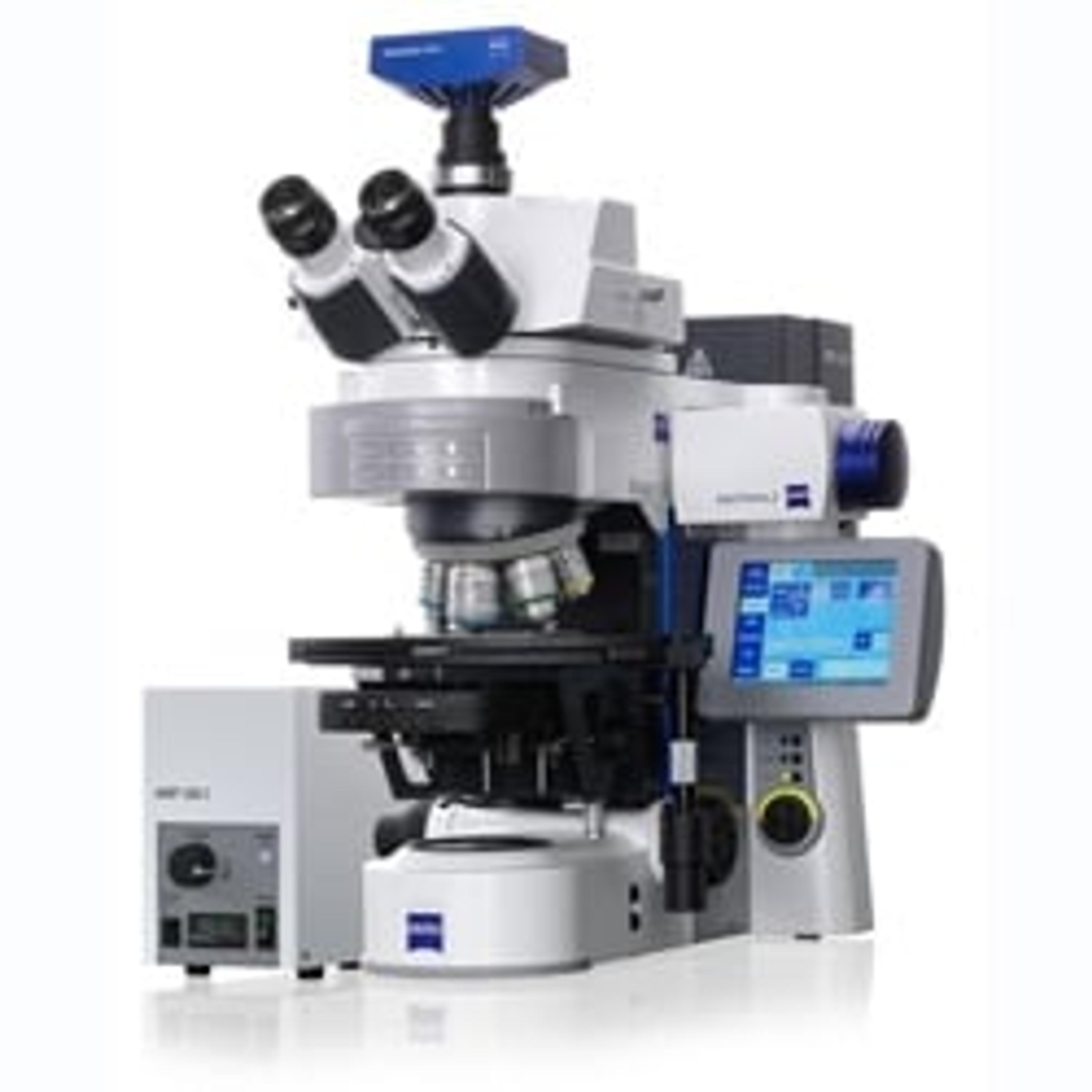
ZEISS ELYRA
ZEISS Research Microscopy SolutionsYour Flexible Imaging System for 3D Superresolution Microscopy.