Biostatus to Showcase Live Zebrafish Imaging HCS at Drug Discovery 2009
31 Aug 2009Product news
CyGEL Sustain now makes it simpler to image zebrafish (Danio rerio) embryos for microscopy, high content screening and for site-specific micro-injections, even from only a few hours post-fertilization. This thermo-reversible mountant is a liquid when cooled below room temperature and sets to a clear, optically-inert gel when warmed. To find out more visit booth D4.
CyGEL Sustain accepts embryo media and anaesthetics such as MS-222, when needed, to maintain yet completely immobilize de-chorionated embryos. After imaging, embryos can be easily recovered with excellent viability even after one hour in CyGEL Sustain thereby permitting observation of further development stages. CyGEL Sustain can equally be used for the mounting of fixed embryos. CyGEL Sustain avoids all the problems associated with methylcellulose and low-melting point agarose.
The far-red DNA dye DRAQ5 remains the counterstain of choice in imaging-based HCS, now complimented by CyTRAK Orange for use on the ArrayScan.
Related products
Request Quote for All Products
CyGEL
Biostatus LimitedA novel thermoreversible gel mountant for temporary immobilization and imaging.
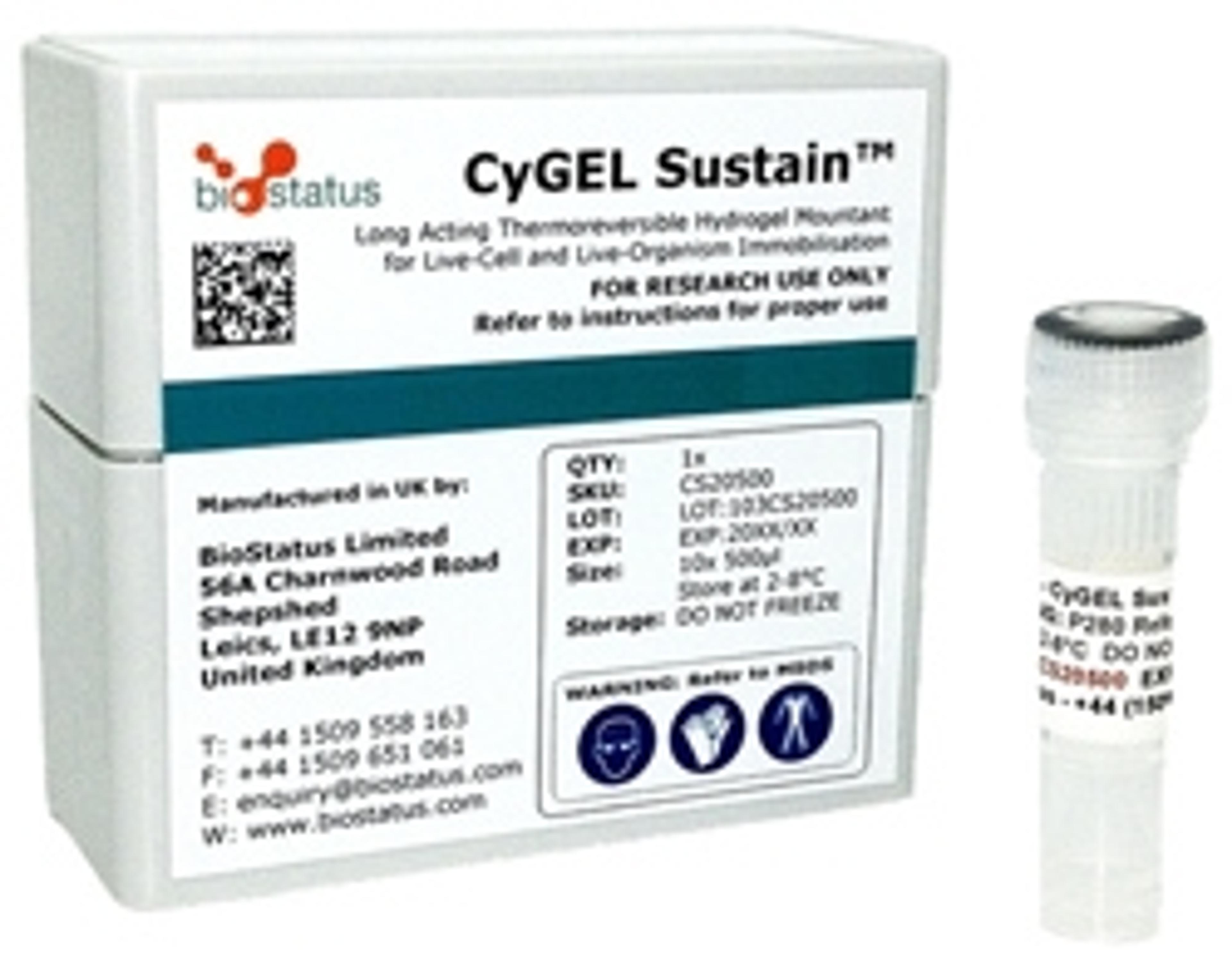
CyGEL Sustain
Biostatus LimitedA novel thermoreversible gel mountant for extended immobilization and imaging.