Automated Microscope for Gentle and Fast Confocal 4D Imaging

Enhancing ZEISS Celldiscoverer 7 with ZEISS LSM 900 for optical sectioning

2 Jul 2019
Georgina Wynne Hughes
Editorial Assistant

Product news

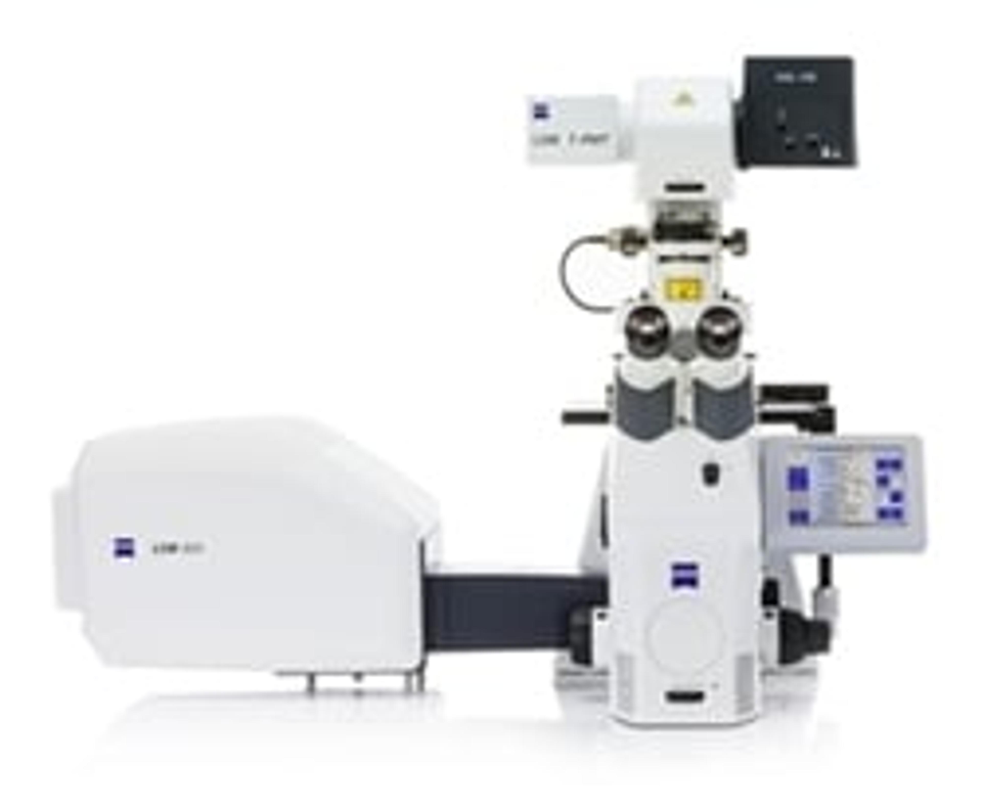
The proven ZEISS Celldiscoverer 7 is a fully integrated high-end imaging system with various incubation and detection options. It combines the easy-to-use automation of a boxed microscope with the image quality and flexibility of a classic inverted research microscope. To get better data from three-dimensional samples, it is now possible to add ZEISS LSM 900 with Airyscan 2 for confocal imaging.

Connecting widefield and confocal – the best of both worlds

Life sciences research often calls for optical sectioning to image samples with best possible contrast and resolution. By adding ZEISS LSM 900 with Airyscan 2 to ZEISS Celldiscoverer 7, users get the ease-of-use and automation from a fully integrated microscope platform and the superb confocal image quality and flexibility of the ZEISS LSM 9 family with Airyscan 2. The new Multiplex mode allows the user to perform super resolution 3D imaging with up to 1.5x higher resolution. Additionally, researchers can easily separate multiple labels with spectral imaging.

A flexible, integrated microscope

ZEISS Celldiscoverer 7 simplifies the laboratory setup and makes work more comfortable. All components are optimized for hassle-free automated imaging. New users and multi-user facilities especially enjoy the in-built automation and usability features when setting up complex experiments. Users can expect better data in shorter times, with less training and maintenance. As the user’s requirements grow, they can expand ZEISS Celldiscoverer 7 with confocal technology, external cameras, deconvolution, and additional environmental control – whatever they need for the challenge of live cell observation.

For more science news, straight to your inbox, join SelectScience today>>

Links

Tags

Light MicroscopyLight microscopes or optical microscopes are used to visualize microscale objects under magnification, including cells, clinical specimens and materials. Lab equipment for light microscopy includes confocal microscopes, fluorescence microscopes, zoom and stereo microscopes. Microscope slides and imaging reagents are available for visualizing samples, as well as various microscope stages and incubators for large or temperature-sensitive samples. Find the best light microscopes in our peer-reviewed product directory: compare products, check customer reviews and receive pricing direct from manufacturers.AutomationAutomation in laboratories and manufacturing processes enhances efficiency, precision, and scalability by reducing the need for manual intervention. It plays a critical role in improving productivity, minimizing human error, and accelerating workflows in fields like diagnostics, drug development, and industrial testing. Automation technologies include robotic systems, automated liquid handlers, and process control systems that streamline complex tasks and ensure consistent, reproducible results. Explore our peer-reviewed product directory to discover the best automation solutions, compare options, read user reviews, and get prices directly from manufacturers.Cell ImagingCell imaging can be achieved using a number of techniques including confocal microscopy, transmission electron microscopy, atomic force microscopy, and light sheet microscopy.ConfocalImagingImaging techniques are essential for obtaining visual representations of samples to understand structures, processes, and function in biological, chemical, and physical research. These tools range from traditional light microscopy to advanced imaging modalities like MRI and electron microscopy, providing researchers with valuable data for diagnostics, drug discovery, and material analysis. Explore imaging solutions in our peer-reviewed product directory to compare products, check reviews, and get pricing directly from manufacturers.Live Cell ImagingLive cell imaging is the study of living cells using microscopes and high-content imaging systems. This technique provides in-depth insight into fast and complex biological processes, by allowing dynamic imaging of living cells instead of acquiring an individual image at a single point in time.MicroscopyMicroscopy is a technique used to observe small objects in detail, from cells to materials, using light or electron microscopes. It enables researchers to examine structures with high resolution, aiding in fields such as biology, medicine, and materials science. With advanced microscopy techniques, scientists can gain insights into cellular processes, tissue structures, and material properties. Explore the best microscopy solutions in our peer-reviewed product directory, compare products, read customer reviews, and get pricing directly from manufacturers.Super Resolution Microscopy
Automated Microscope for Gentle and Fast Confocal 4D Imaging