A Major Step Forward for Structural Biology and Drug Discovery
Thermo Fisher Scientific and Diamond Light Source open industrial access to largest cryo-electron microscopy sites in the world
12 Sept 2018
Industry news

An agreement to launch a new cryo-EM capability for use in the life sciences industry sector by Thermo Fisher Scientific, one of the world leaders in high-end scientific instrumentation, and Diamond Light Source, the UK’s national synchrotron and one of the most advanced scientific facilities in the world, was announced at the official opening of the new national electron bio-imaging centre (eBIC) at Diamond.
This announcement confirms Diamond as one of the major global cryo-EM sites embedded with an abundance of complementary synchrotron-based techniques, and thereby, provides the life sciences sector with an offer not available anywhere else in the world.
Professor Dave Stuart, Life Sciences Director at Diamond and MRC Professor of Structural Biology at the University of Oxford, Department of Clinical Medicine, says, "Access to 21st century scientific tools to push the boundaries of scientific research is essential for both academia and industry, and what we have created here at Diamond is truly unique in the world in terms of size and scale. The new centre offers the opportunity for almost real-time physiology, capturing proteins in action at cryo-temperatures by flash-freezing them at various stages. What Diamond has created with eBIC is an integrated facility for structural biology, which will accelerate R&D for both industry and academic users. The additional advanced instruments made available by Thermo Fisher will position the UK as a global leader in providing large-scale industrial access to cryo-EM for drug discovery research. Our new collaboration provides a step change in our offer for industry users and helps ensure that R&D remains in the UK.”
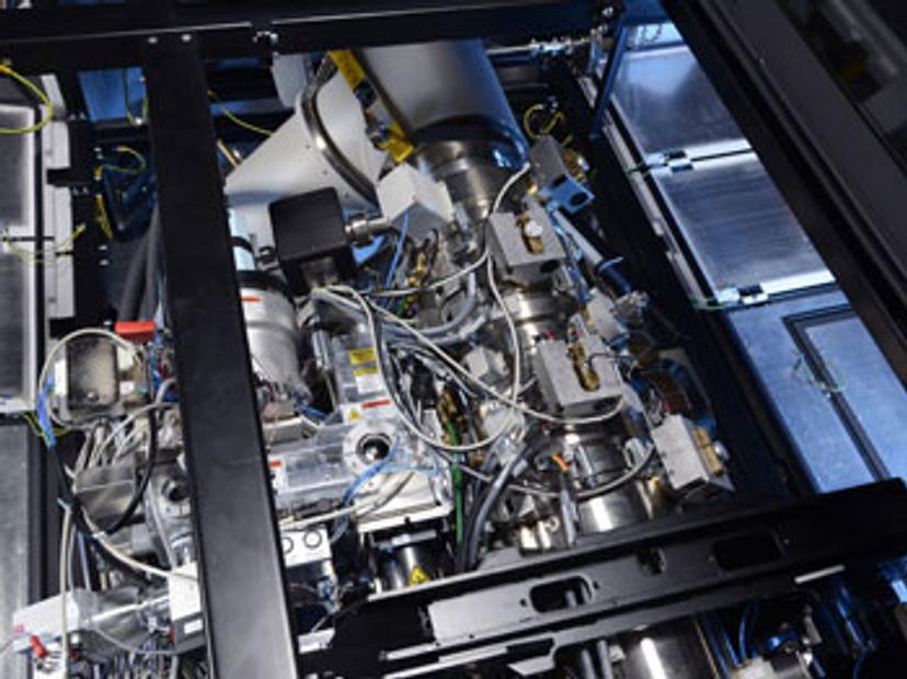
The collaboration with Thermo Fisher will further expand Diamond’s cryo-EM offerings by providing two new dedicated microscopes and professional cryo-EM services designed exclusively for the pharmaceutical industry. New instruments being installed include a Thermo Scientific Glacios Cryo Transmission Electron Microscope (TEM) and a Thermo Scientific Krios Cryo-TEM at eBIC. The two organisations will provide cryo-EM guidance and expertise in sample preparation, cryo-EM sample screening, and high-end data collection to industrial researchers.
It is an area of huge interest to the pharmaceutical industry because we are able to visualise novel atomic structures of new drug targets and, at the same time improve our understanding of challenging current targets.
Dr. John Barker Senior Vice President at Evotec
“Researchers at leading pharmaceutical companies are already using instruments from Thermo Fisher Scientific to drive impactful research that can help speed the path to understanding and treating viruses and diseases,” says Mike Shafer, President, Materials and Structural Analysis, Thermo Fisher. “The combination of the expert staff members at eBIC and Diamond, alongside these new Thermo Scientific cryo-EM instruments, ensures these researchers are on the cutting edge of discovery.”
The new microscopes will add to Diamond’s four existing high-end microscopes, which are starting operation within eBIC and will continue to support the academic demand.
Recent technology developments in cryo-EM are proving to be powerful for both basic and applied science at Diamond. This is why Diamond, with the wider community led by Birkbeck College and Oxford University, established a centre embedded within the synchrotron's infrastructure through a strategic grant from the Wellcome Trust, Biotechnology and Biological Research Council and Medical Research Council. The centre officially opens today by Dr Richard Henderson from the Laboratory of Molecular Biology, Cambridge. Dr Henderson is one of the recipients of the 2017 Nobel Prize for Chemistry for developing the cryo-EM technique.
Dr Henderson says: “Using single particle cryo-EM, researchers can now visualise biomolecules at near atomic resolution. This technology has taken biochemistry into a new era, allowing atomic structure determinations of many protein and other macromolecular building blocks that were previously very difficult or impossible to obtain. The technique also opens up high resolution studies of macromolecules in their cellular context through the use of electron cryotomography. Coupling these techniques with the capabilities at Diamond creates a unique environment that will help keep the UK at the forefront of world-leading science.”
Structural insight into proteins, viruses and other macromolecular complexes helps pharmaceutical companies better understand disease mechanisms, more quickly assess possible drug targets, and accelerates optimisation of these drug targets. Mike Shafer adds: “It’s important for Thermo Fisher to support this effort with both equipment and staffing, allowing researchers to maximize their time spent with the instruments. We are excited Diamond will provide joint staff, expertise in swift and effective data collection and analysis, and support with sample preparation.”
Access to 21st century scientific tools to push the boundaries of scientific research is essential for both academia and industry, and what we have created here at Diamond is truly unique in the world in terms of size and scale
Professor Dave Stuart Life Sciences Director at Diamond
Pharmaceutical company researchers will have easy access to expert knowledge and industry leading cryo-EM equipment, covering the complete workflow for single particle analysis in the first instance. Access to both a Glacios Cryo-TEM and a Krios Cryo-TEM at one location will optimise productivity and time-to-result because of the designed-in connectivity between the two instruments. Scientists will be able to first pre-screen samples on the Glacios Cryo-TEM to find the best quality samples before advancing to the higher resolution imaging on the Krios Cryo-TEM.
The infrastructure on offer at eBIC taps into the expertise of Diamond as a 24-hours per day, six days a week operational model. Typically, eBIC industry users are granted one to three day sessions depending on their requirements. The two new microscopes will have four staff providing support for year-round operation.
Early users of the facility comments include:
Dr John Barker, Senior Vice President and Global Head of Protein Sciences at Evotec says: “The area of single particle cryo-EM is a rapidly developing technology that is delivering a new approach to looking at previously intractable structural questions. It is an area of huge interest to the pharmaceutical industry because we are able to visualise novel atomic structures of new drug targets and, at the same time improve our understanding of challenging current targets. The decision to expand eBIC, with a focus on industrial users, is an exciting opportunity for UK-based industry to continue to be at the forefront of structural biology in drug discovery.”
Dr Gillian Burgess, Site Head & VP, Research at Vertex Pharmaceuticals Oxford says:
“We have collaborated with Diamond Light Source since its inception 10 years ago. The announced investment in cryo-electron microscopy at the Harwell Campus will transform access to this innovative technology for the Life Sciences Industry. We will be able to image large and complex proteins in a way that has previously not been possible with more traditional techniques. At Vertex we are committed to the development of new precision medicines that can treat serious diseases and we believe that cryo-electron microscopy can accelerate the discovery of such medicines.”