89 North Introduce the Heliophor™ Fluorescence Illumination System
20 Apr 2011Product news
The Heliophor from 89 North, Inc. is a pumped-phosphor, fluorescence illumination system designed for a wide range of fluorescence imaging applications including:
• Multichannel live cell imaging
• FRET
• High content screening
• Quantitative fluorescence imaging
• High speed, ratiometric imaging
The pumped-phosphor design has all of the advantages of conventional solid state illumination systems, including high speed digital switching, stable output intensity and extremely long lifetimes. However, where other solid state illumination systems have lower intensity in the green and red regions of the spectrum, the Heliophor has excellent output in those and also in other wavelength regions.
The Heliophor comes equipped with a host of standard features that others charge for as “extras”. These include:
User exchangeable modules
Up to 6 modules can be installed per system
Ideal for core facilities
Easy field installation/exchange of modules by user
Easily reconfigure system for different experimental protocols
For more information, visit the company article page.
Related products
Request Quote for All Products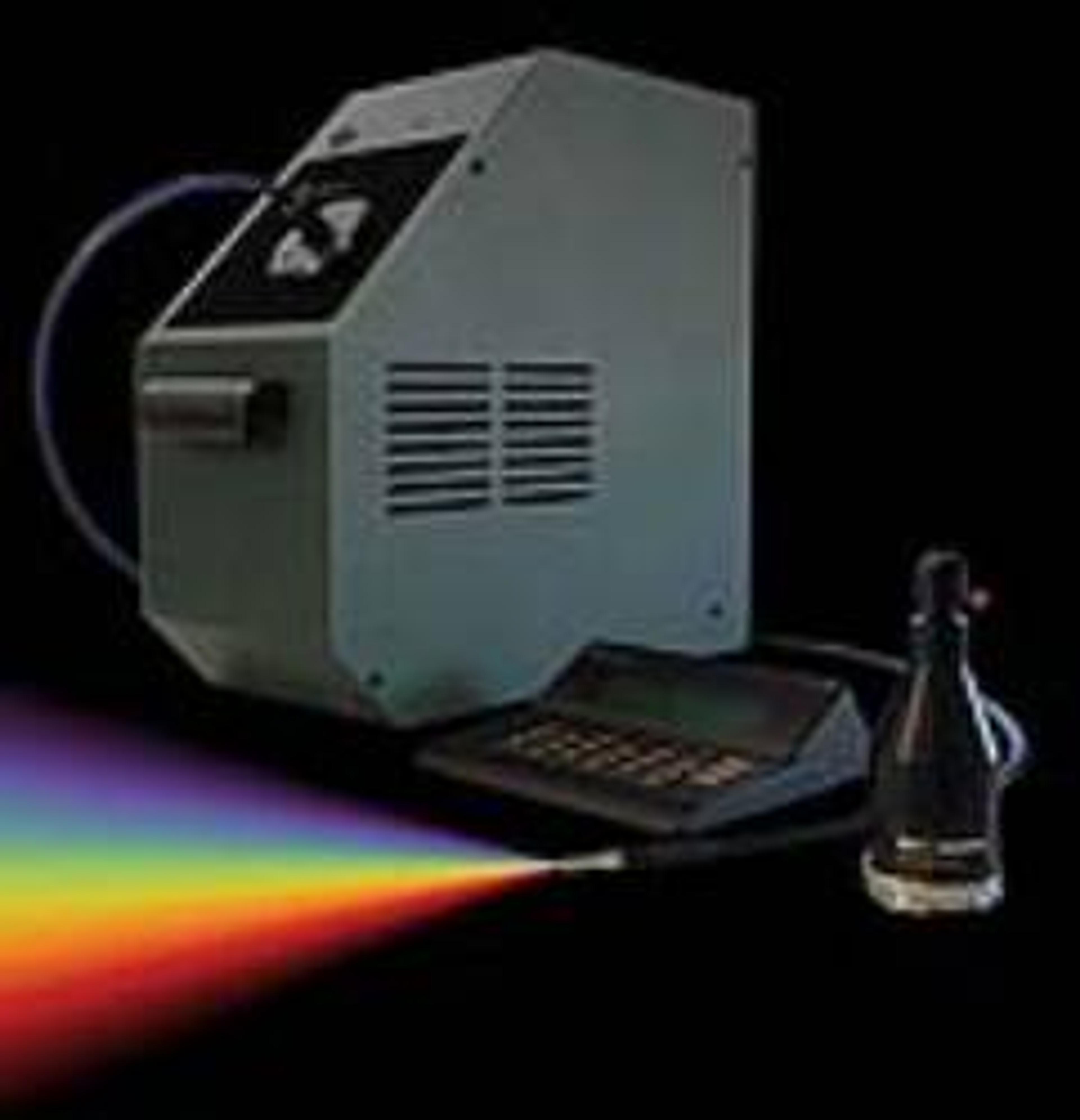
Heliophor
89 NorthHeliophor Light Engine 89 North's Heliophor, is a pumped phosphor light engine for quantitative fluorescence imaging. It includes up to 6 user-exchangeable wavelength modules and features rise and fall times less than 10 microseconds. The Heliophor's digital shuttering and onboard macro capabilities enable high-speed, multidimensional imaging. Backed by a limited lifetime warranty, the Heliophor delivers consistently high intensity, ultra-stable output.