10 Scientists Leading the Fight Against Cancer

SelectScience celebrates scientists at the forefront of cancer research, from liquid biopsies through cancer stem cells to drug delivery innovations

2 Feb 2018
Frankie MacDonald
Administrator / Office Personnel

Editorial article

Sunday, February 4, marks the internationally recognized ‘World Cancer Day’, an opportunity for people worldwide to raise awareness and unite in the battle against cancer. These 10 scientists, from across the globe, employ diverse technologies such as live-cell imaging, optical methods, MRI, sequencing, among others, all leading to the same destination – to emerge as winners in the tough fight against the disease. We salute their efforts.

1. Researchers Deplete Cancer Stem Cells to Fight Recurring Brain Tumours

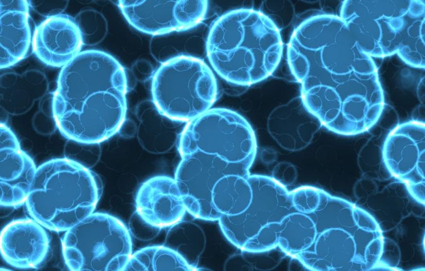
Dr. Raffaella Spina, of Case Western Reserve University, focuses her research on tackling glioblastoma multiforme, an aggressive stage IV brain cancer associated with poor patient outcome. Dr. Spina and colleagues investigate controlling the fate of stem cells (SCs) responsible for propagating these tumors, such as forcing differentiation into non-cancerous astroglial progeny. Their most recent research also involved disruption of the MCT-4 transporter, compromising SC survival and tumor proliferation as a result. Read more

H&E staining of a brain section showing invasive and proliferative tumours Image courtesy of Dr. R Spina

2. How Retrospective Genetic Studies at NIH Are Providing Answers for the Future of Cancer

Genetic mutations and genomic instability are at the core of tumor development. If we can recognize patterns in patient populations that allow us to identify driver mutations, then we can use this information to develop novel therapeutics. Dr. Stephen Chanock, Director of Cancer Epidemiology and Genetics at the U.S National Cancer Institute, NIH, conducts large-scale, retrospective, genome-wide sequencing studies with an overarching goal of mapping susceptibility alleles in primary and metastatic cancers. Read our exclusive Q & A with Dr. Chanock.

Samples from international cancer patient cohorts enable retrospective genome wide studies Image courtesy of Ed Uthman under the Creative Commons License

3. Detecting Genetic Mutations in Bladder Cancer Patients Using Droplet Digital PCR

With technological advances, the ability to detect circulating tumor DNA in so-called ‘liquid biopsies’ has become possible. Watch this video interview with Emil Christensen, a Ph.D. student from Aarhus University, Denmark, in which he discusses the value of using blood and urine biopsies for predicting the metastatic potential of non-invasive primary bladder cancers, by assessing the quantity and type of genetic mutations in sample fragments. Watch the video

Emil Christensen, of Aarhus University, Denmark

4. Continuous Live Imaging Reveals Why Some Prostate Cancer Cells Resist Androgen-Deprivation Therapy

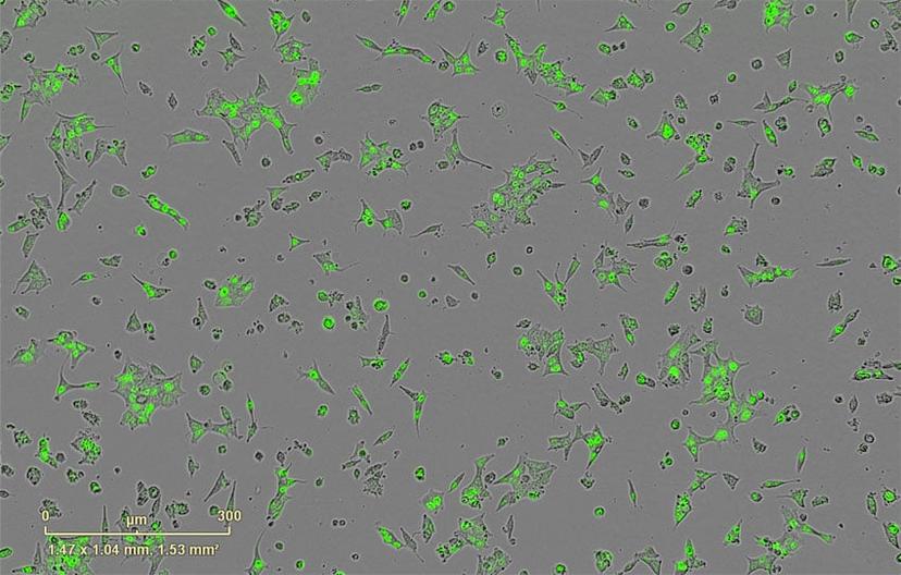
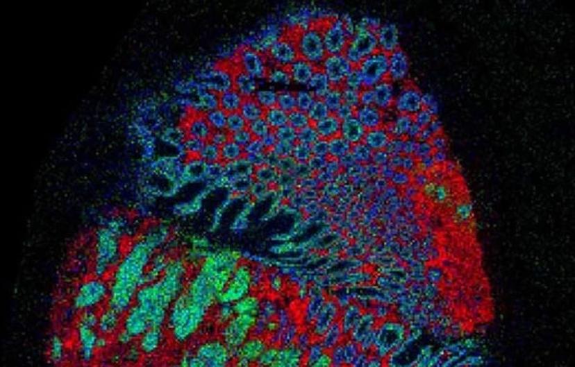
Live-cell imaging is a powerful platform for basic and translational research. Using this technology we can further our understanding of important sub-cellular processes and interactions. Fiorella Magani, of Miami University, utilizes fluorescent labeling in real time to understand the molecular basis of castrate-resistant prostate cancer (CRPC). Read this article to find out more about her specific research into splice variants of the androgen receptor, often upregulated in CRPC.

Human castration-resistant cancer cell line 22Rv1 fluorescently labeled with NucLight Green BacMan Image courtesy of Fiorella Magani.

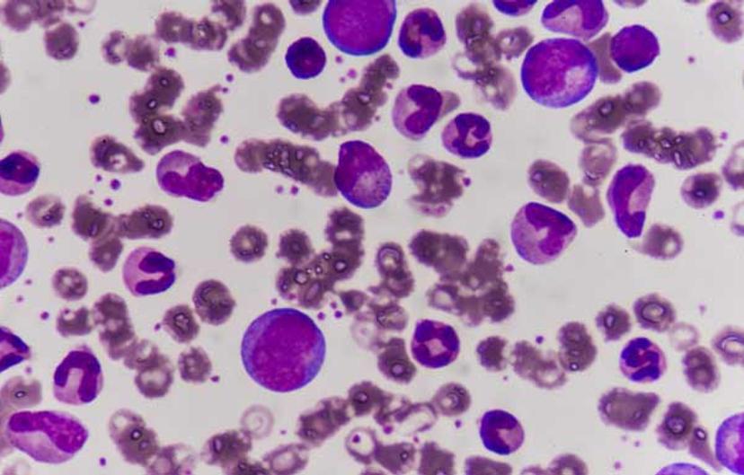
5. Advances in CAR T Therapy: Reprogramming Patient Cells to Treat Cancer

CAR T, chimeric antigen receptor modified T cell therapy, is an innovative approach in treating cancer, that involves genetic reprogramming of patient immune cells to both recognize and attack cancerous B cells. Read this SelectScience interview with Dr, Richard Jones, of Beckman Coulter, to find out more about the promise of CAR T in treating advanced stage disease.

CAR T therapy offers promise for treatng leukemia image source: chirawan Somsanuk © 123RF.com

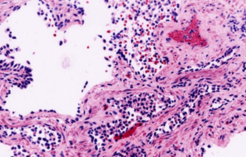
6. New Optical Method “Cuts” and Examines Cancer Biopsies During Surgery

Surgical tumor resection is a key component of cancer therapy for many patients. However, it is important that, where possible, the surgeon excises the tumor with an appropriate margin to ensure no cells are left behind. Dr. Quincey Brown, of Tulane University, and colleagues, have developed novel optical approaches for prostate cancers that will prevent damage to adjacent structures and reduce the likelihood of surgical spill which often encourages recurrence. Read this SelectScience interview with Dr. Brown to find out more.

Prostate cancer tissue stained for DRAQ5TM and eosin

7. Dr. Maria Lambros; In vivo imaging to Optimize of Liposome-Mediated Drug Delivery in Cancer

Honing drug delivery to target cancer cells exclusively, is a hugely important area of cancer research. Not only does it improve drug efficacy, but also reduces toxicity to non-cancerous tissues. In an interview with Dr. Maria Lambros, SelectScience finds out more about her work with liposome-encapsulated drugs with live in vivo imaging for kinetic analysis.

Live in vivo imaging for measuring mouse xenograft pancreatic tumour growth (Image courtesy of Analytik Jena.)

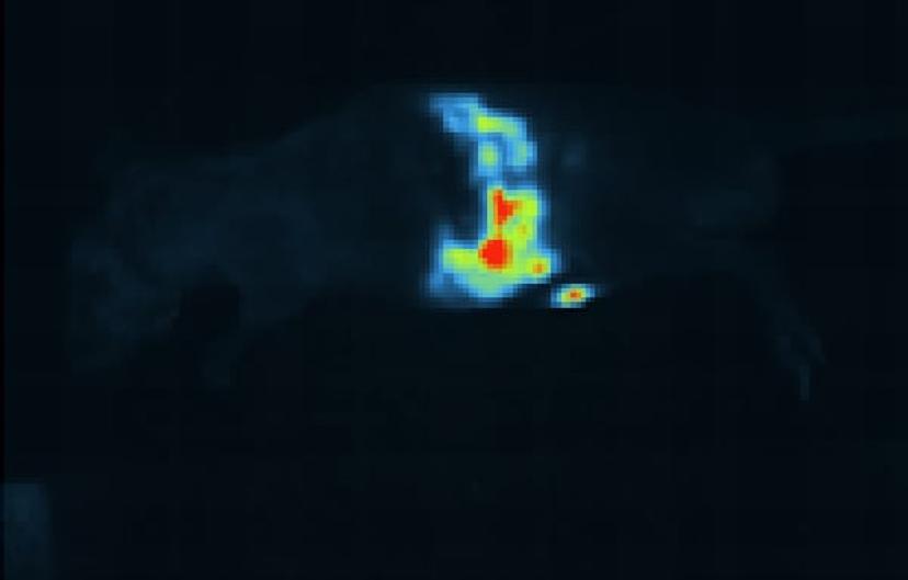
8. A 3-Dimensional ‘Google Earth’ Style Map for Tumors

Dr. Josaphine Bunch, of the National Physics Laboratory, London, employs a combination of mass spectrometry techniques to map tumors at the gross and molecular level, in 3D. This technology can also be used to differentiate tumor from non-tumor tissue and will enable us to understand more of the molecular and metabolic basis of cancers for delivering targeted therapy. Read the interview

Bowel Cancer image assembled using multiple mass spectrometry techniques Image credit: Zoltan Takats, Renata Filipe-Soares (Imperial College London); Nicole Strittmatter, Gregory Hamm, Richard Goodwin (Astra Zeneca); Rory Steven, Adam Taylor, Alan Race, Spencer Thomas, Rasmus Havelund, Josephine Bunch (NPL)

9. 3D Bioengineered Brains – a New Approach to Personalized Cancer Therapy

In pre-clinical trials, it is vital to analyze drugs in cancer models that most accurately represent the disease in humans. Dr. Min Tang-Schomer is working at the forefront of the tissue bio-engineering field, having developed brain-mimicking tissue that models grey and white matter in composite, with electrophysiological features. Read this article to find out about her current research that involves developing brain models derived from patient-extracted cells for a personalized medicine approach.

Source: 123rf.com 2014

10. Cancer Biologists Investigate Epigenetic Regulation of Pancreatic Cancer Cells using Live Cell Imaging

Epigenetic regulation of the genome is equally important as the information within our DNA. In this interview, cancer researchers from the MD Anderson Cancer Center, Dr. Sonal Gupta and Dr. Sammy Ferri-Borgogno, give SelectScience an insight into their work on epigenetic regulators in pancreatic cancer, one of the most aggressive cancer diagnoses.

Links

Tags

Mass SpectrometryMass spectrometry (MS) is a powerful analytical technique used to identify and quantify molecules based on the mass-to-charge ratio of gas-phase ions. It provides detailed information about the structure, composition, and properties of compounds and is widely used across fields such as environmental monitoring, materials science, drug discovery and development, food and beverage testing, and wider chemical research. Key MS techniques include tandem mass spectrometry (MS/MS), liquid chromatography–mass spectrometry (LS-MS) and inductively coupled plasma (ICP-MS). Choosing from these wide range of techniques and technologies can be a daunting task, so keep up to date with scientific applications, performance expectations, and customer reviews here all in one place. Visit our product directory to receive quotes direct from the manufacturer. Cell / Tissue CultureCell culture or tissue culture is used to study the biology of cells or tissues and to isolate cellular products in an environment which can be manipulated and well defined. Accurately control your culture environment with bioreactors or culture incubators, bind your cells to a surface or together with an extracellular matrix. Distinguish cell types with differential media or proliferate cells with certain characteristics using selective media. Enrich your media with supplements such as growth factors, sera and vitamins. Find the best cell and tissue culture products, kits and equipment in our peer-reviewed product directory: compare products, check customer reviews and receive pricing direct from manufacturers.Cell-Based AssaysCell-based assays are used to monitor the presence, quantity and activities of a desired cellular analyte including drug molecules or biomarkers. This can reveal information on cell health (apoptosis, cytotoxicity, viability and proliferation assays), cell metabolism, cell migration and cell signaling mechanisms. Find the best cell-based assay products, kits and equipment with our peer reviewed product directory: compare products, check customer reviews and receiving pricing direct from manufacturers.Gene Expression and Molecular CloningMolecular cloning is a set of techniques that utilizes vectors to transfer recombinant DNA into host cells and is an essential tool for investigating the expression of genes and proteins in bacterial or mammalian cells. A variety of vectors optimized for gene cloning and expression in a range of host organisms are available, alongside competent cells for genetic replication. Here, you can explore a range of molecular tools, high-quality genomic and cDNA libraries, premade clones, transformation and transfection reagents and mutagenesis or gene expression detection assays and expression arrays. Find the best gene expression and molecular cloning products in our peer-reviewed product directory: compare products, check customer reviews and receive pricing direct from manufacturers.In Vivo Imaging Systems<i>In vivo</i> imaging systems, including pre-clinical imaging systems and medical imaging systems are used to non-invasively visualize and capture images of live animals and plants. Monitor the natural processes or diseases of your subjects using small-animal pre-clinical imaging systems, including single photon positron emission tomography (SPECT), positron emission tomography (PET), computed tomography (micro-CT), magnetic resonance imaging (MRI), X-ray radiography, ultrasound, fluorescence and bioluminescence imagers. Multimodal systems and software solutions are also available for correlative analysis of organ, tissue, cell, or molecular-level processes. Find the best in vivo imaging products in our peer-reviewed product directory: compare products, check customer reviews and receive pricing direct from manufacturers.Cancer ResearchCancer research aims to understand the mechanisms of cancer development and progression to improve prevention, diagnosis, and treatment. From molecular biology to clinical trials, research spans a wide range of disciplines, including immunotherapy, targeted therapies, and drug discovery. Explore the best cancer research products in our peer-reviewed product directory; compare products, check reviews, and get pricing directly from manufacturers.Protein BiologyThe analysis of protein expression, identity and function is vital for many areas of life science research and drug discovery. Some of the most commonly used techniques in protein analysis include Western blotting, electrophoresis and mass spectrometry.